Amygdala Drawing
Amygdala Drawing - Hippocampal sulcus remnant cysts seen on t2 axial mri (magnetic resonance imaging) human brain, illustration. Click now to learn more at kenhub! Amygdala medical labeled vector illustration and scheme with. Although we often refer to it in the singular, there are two amygdalae —one in each cerebral hemisphere. Latin from greek, ἀμυγδαλή, amygdalē, 'almond', 'tonsil') is a paired nuclear complex present in the cerebral hemispheres of vertebrates. Adapting practices for students with special needs. The amygdala is a complex grey matter structure located anterior and superior to the temporal horn of the lateral ventricle and head of the hippocampus. How to draw human brain. Give a simple definition of the amygdala, the hippocampus and prefrontal cortex. It is part of the limbic system and plays a key role in processing emotions and emotional reactions. 11k views 6 years ago. Amygdalae) is a very well studied part of the limbic system and forms part of the mesial temporal lobe. Check out amazing amygdala artwork on deviantart. O the names of these parts of the brain are the prefrontal cortex,. Amygdala medical labeled vector illustration and scheme with. Latin from greek, ἀμυγδαλή, amygdalē, 'almond', 'tonsil') is a paired nuclear complex present in the cerebral hemispheres of vertebrates. It's a key part of emotional control and processes. The amygdala is a complex structure of cells nestled in the middle of the brain, adjacent to the hippocampus (which is associated with memory formation). Web this article describes the anatomy of. Web how to draw human brain amygdala anatomy drawing. The colour of the amygdala is that of the grey substance. The role of the amygdala. Web identify the amygdala, the hippocampus, and prefrontal cortex on a diagram of the brain. Although we often refer to it in the singular, there are two amygdalae —one in each cerebral hemisphere. Hippocampal sulcus remnant cysts seen on t2 axial mri (magnetic resonance imaging) human brain, illustration. Amygdala of the brain, artwork. Web on every picture of the artwork, the amygdala is surrounded by a circle so as to be displayed well. Point to the prefrontal cortex, the hippocampus, and the amygdala and explain: Apply their new knowledge of the brain to. Web want to discover art related to amygdala? Web the amygdala (/ ə ˈ m ɪ ɡ d ə l ə /; The amygdala is a complex grey matter structure located anterior and superior to the temporal horn of the lateral ventricle and head of the hippocampus. Latin from greek, ἀμυγδαλή, amygdalē, 'almond', 'tonsil') is a paired nuclear complex present. Click now to learn more at kenhub! The amygdala is a complex structure of cells nestled in the middle of the brain, adjacent to the hippocampus (which is associated with memory formation). The colour of the amygdala is that of the grey substance. Web on every picture of the artwork, the amygdala is surrounded by a circle so as to. The colour of the amygdala is that of the grey substance. Point to the prefrontal cortex, the hippocampus, and the amygdala and explain: How to manage amygdala hijack. It is considered part of the limbic system. Amygdala medical labeled vector illustration and scheme with. The amygdala is a paired structure (the two are considered one brain area) inside your temporal lobe. It is part of the limbic system and plays a key role in processing emotions and emotional reactions. The colour of the amygdala is that of the grey substance. It also plays a role in memory and learning. O the names of these. Web mindful activity (15 minutes) • focus the children’s attention to the large drawing of the brain. Web identify the amygdala, the hippocampus, and prefrontal cortex on a diagram of the brain. Hippocampal sulcus remnant cysts seen on t2 axial mri (magnetic resonance imaging) human brain, illustration. The colour of the amygdala is that of the grey substance. Web drawing. Web schematic drawing of the human brain, indicating the location of amygdala from a coronal perspective. Get inspired by our community of talented artists. Adapting practices for students with special needs. Although we often refer to it in the singular, there are two amygdalae —one in each cerebral hemisphere. How to draw human brain. Amygdala of the brain, artwork. Amygdalae) is a very well studied part of the limbic system and forms part of the mesial temporal lobe. Web the amygdala (/ ə ˈ m ɪ ɡ d ə l ə /; Web want to discover art related to amygdala? Check out amazing amygdala artwork on deviantart. It's a key part of emotional control and processes. Point to the prefrontal cortex, the hippocampus, and the amygdala and explain: Click now to learn more at kenhub! Segmentation the amygdala is segmented using a contour line and manual editing. How to draw human brain. Apply their new knowledge of the brain to everyday scenarios. Adapting practices for students with special needs. The amygdala is a complex structure of cells nestled in the middle of the brain, adjacent to the hippocampus (which is associated with memory formation). Amygdala hijack and mental health. The role of the amygdala. The amygdala is a complex grey matter structure located anterior and superior to the temporal horn of the lateral ventricle and head of the hippocampus.
PSYCH 260

Pin on La photographie

Brain amygdala anatomy Royalty Free Vector Image

And as you’ve valiantly hunted high and low for answers to this
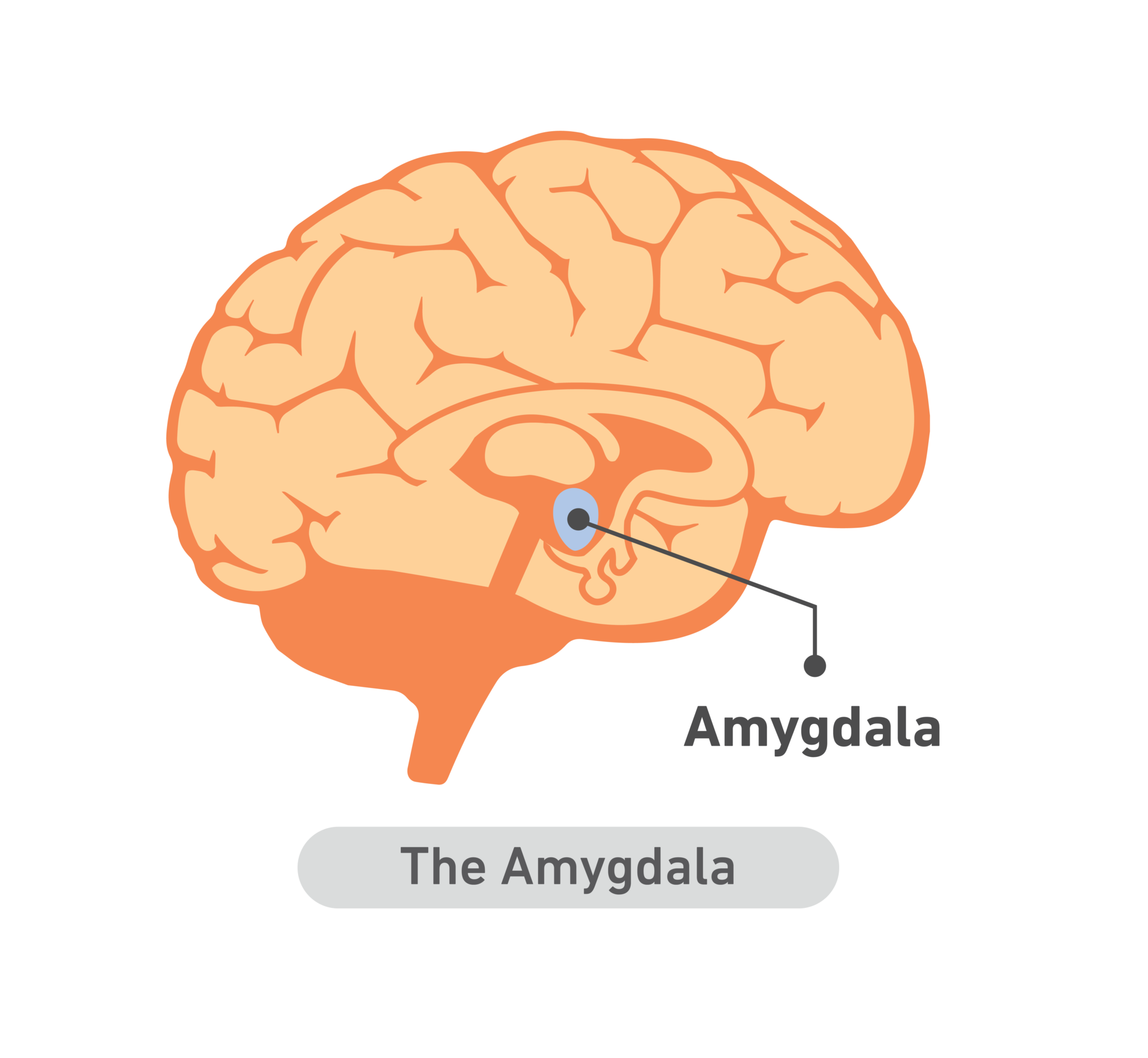
The amygdala The Field Guide
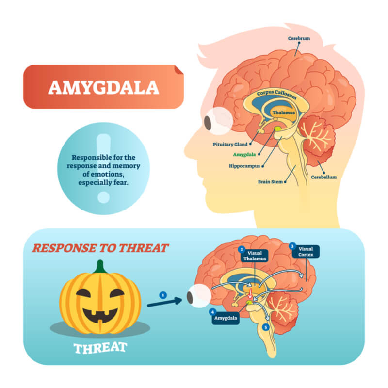
Limbic System And Emotion
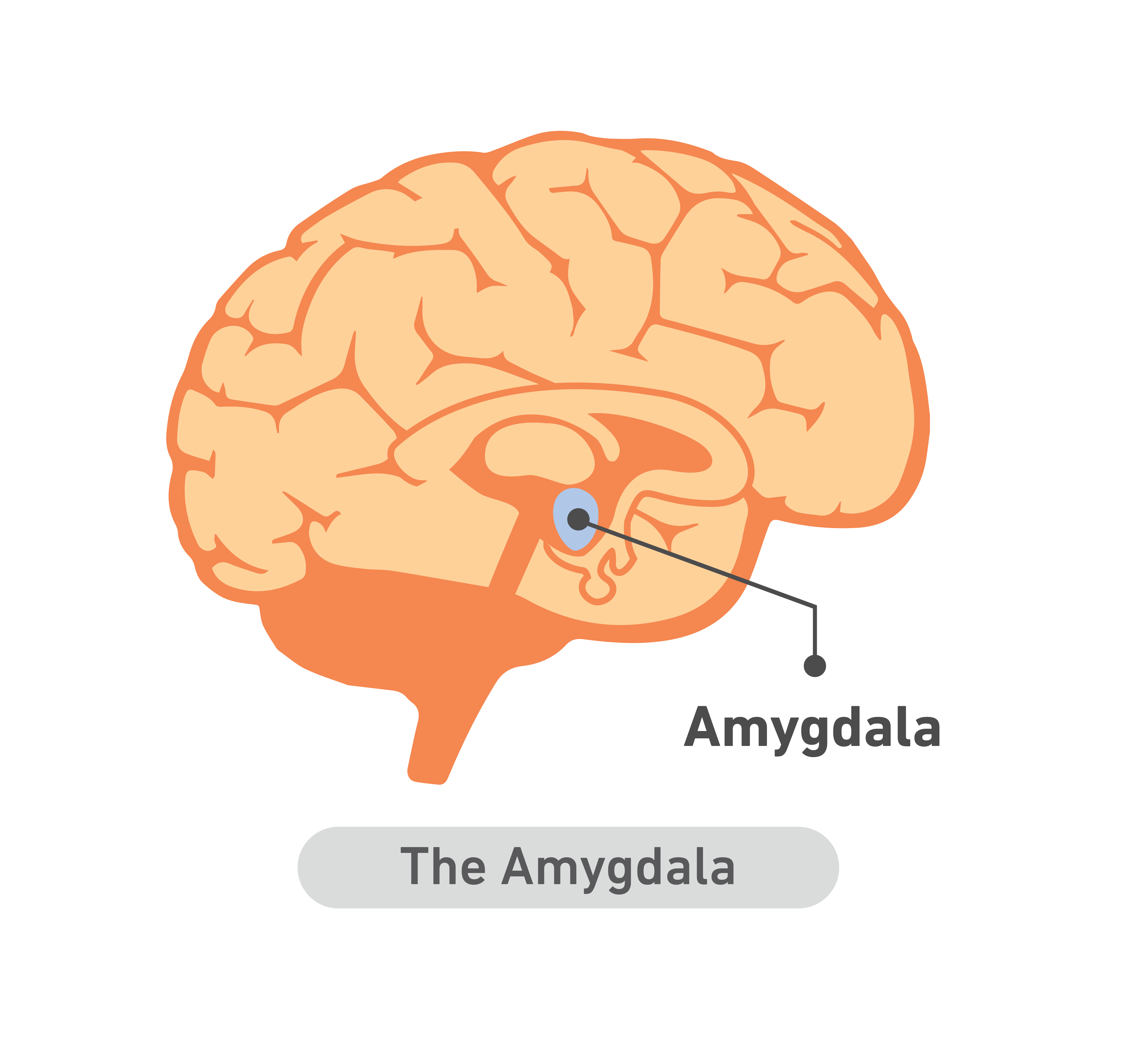
The amygdala The Field Guide
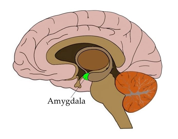
Know Your Brain Amygdala
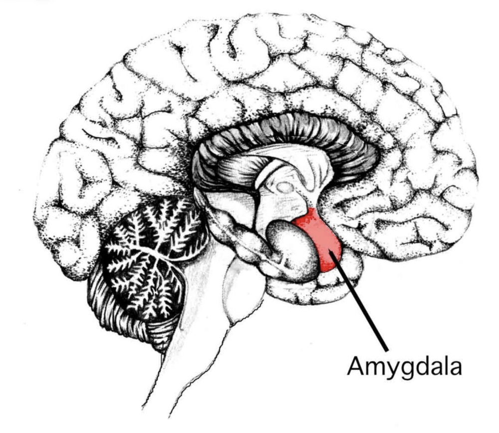
amygdala
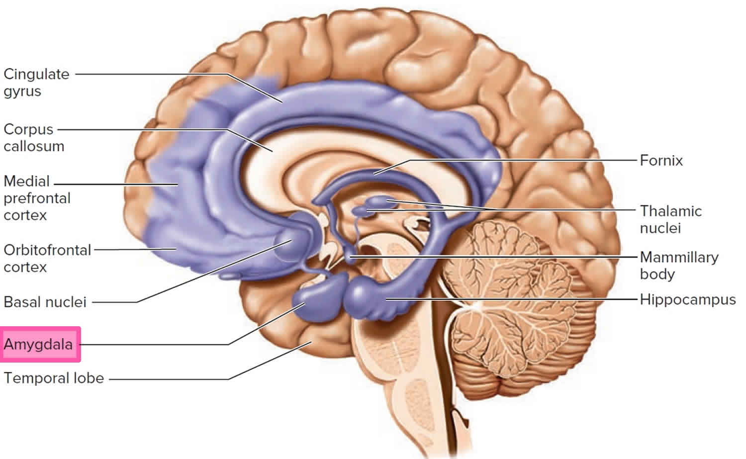
Amygdala function, location & what happens when amygdala is damaged
The Amygdala Is A Paired Structure (The Two Are Considered One Brain Area) Inside Your Temporal Lobe.
Amygdala Medical Labeled Vector Illustration And Scheme With.
Web The Amygdala Is A Collection Of Nuclei Found Deep Within The Temporal Lobe.
Although We Often Refer To It In The Singular, There Are Two Amygdalae —One In Each Cerebral Hemisphere.
Related Post: