Cartilage Drawing
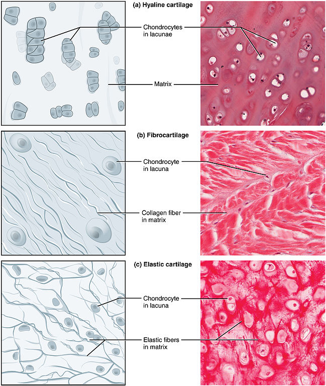
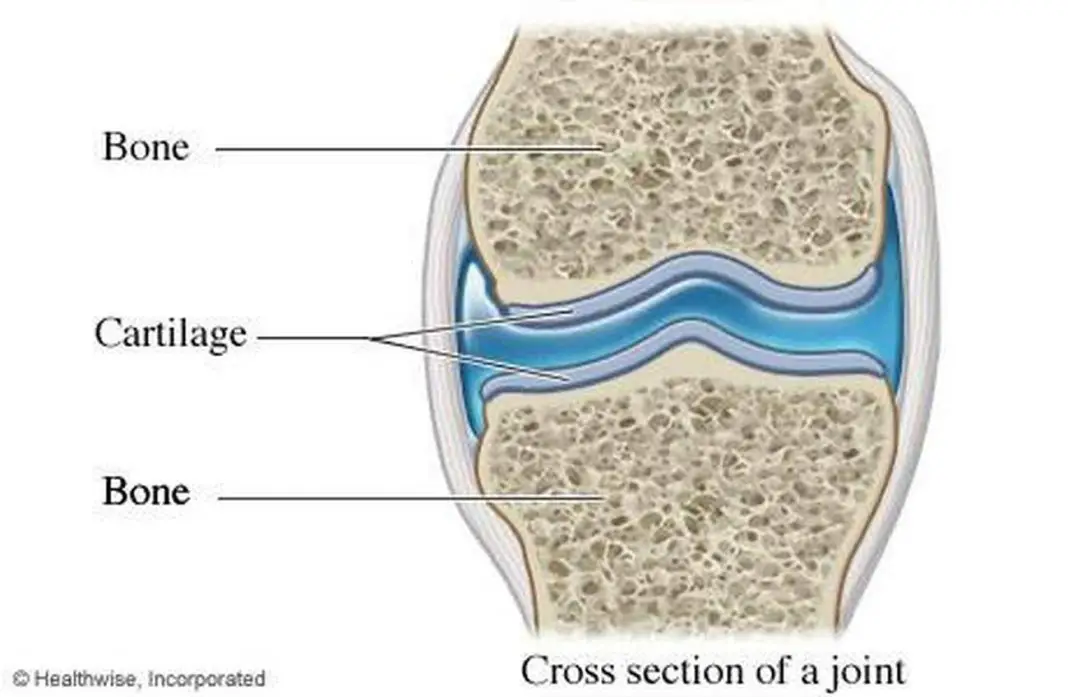
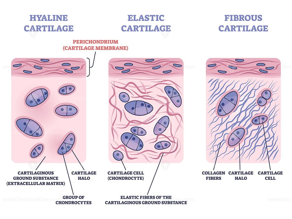
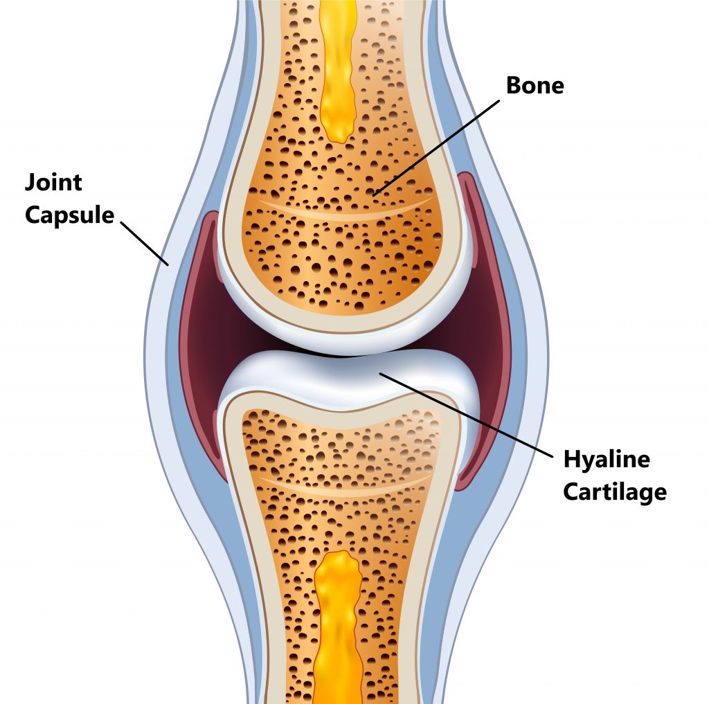
Cartilage Drawing - Web this result snaps a four match losing streak, and this combined with newcastle’s draw means they only need one point from their last two games (at city, home to sheffield united) to clinch fifth. First, i would like to point out the essential histological features from the hyaline cartilage histology slide under the light microscope. Hyaline cartilage, the most abundant type of cartilage, plays a supportive role and assists in movement. Cartilage is a connective tissue composed of chondrocytes (cells) that produce long proteins called fibers, and smaller molecules such as glycoproteins that are collectively referred to as ground substance. Web chapter 1 cartilage morphology. Web elastic cartilage, sometimes referred to as yellow fibrocartilage, is a type of cartilage that provides both strength and elasticity to certain parts of the body, such as the ears. Hyaline cartilage hyaline cartilage is the most widespread cartilage type and, in adults, it forms the articular surfaces of long bones, the rib tips, the rings of the trachea, and parts of the skull. Web cartilage, bone and bone development. Isogenous groups and interstitial growth results when chondrocytes divide and produce extracellular matrix. Territorial matrix lies immediately around each isogenous group and is high in glycosaminoglycans. Step by step drawing of elastic cartilage how to draw elastic cartilage, histology journal.more. Web during embryonic development, hyaline cartilage serves as temporary cartilage models that are essential precursors to the formation of most of the axial and appendicular skeleton. Therefore, this matrix stains more intensely than matrix farther from an isogenous group, the interterritorial matrix. Fetal face, frontal section,. It contains polysacchride derivaites called chondroitin sulfates which complex with protein in the ground substance forming proteoglycan. Hyaline cartilage provides support and flexibility to different parts of the body. Ear pinna, aldehyde fuchsin and masson, 20x (elastic cartilage). First, i would like to point out the essential histological features from the hyaline cartilage histology slide under the light microscope. Cartilage. Cartilage tissue is avascular and therefore relies on obtaining its nutrients via diffusion, sometimes even over large distances. It contains polysacchride derivaites called chondroitin sulfates which complex with protein in the ground substance forming proteoglycan. Hyaline cartilage is high in collagen, a protein that is found not only in connective tissue but also in skin and bones, and helps hold. Ac is a dense connective tissue mainly comprised of collagen, proteoglycans, organized in special zones containing special types of cells called articular chondrocytes [ 1, 2 ]. Step by step drawing of histology of hyaline cartilage Cartilage is a connective tissue composed of chondrocytes (cells) that produce long proteins called fibers, and smaller molecules such as glycoproteins that are collectively. Hyaline cartilage, fibrocartilage, and elastic cartilage. Epiglottis, h&e, 20x (elastic cartilage). The four groups were evaluated by assessing tissue regeneration within the defect. Ear pinna, aldehyde fuchsin and masson, 20x (elastic cartilage). A type of cartilage that is characteristically glossy and smooth in appearance, and with interstitial substance containing fine type ii collagen fibres obscured by the ground substance. Epiglottis, h&e, 20x (elastic cartilage). Web likecomment share subscribe #hyalinecartilage #histodiagrams #hyalinecartilagediagram #cartilagehistology Web matrix of hyaline structure. 2.9k views 4 years ago vadnagar. Cartilage tissue is avascular and therefore relies on obtaining its nutrients via diffusion, sometimes even over large distances. Decreases friction and distributes loads. Web articular cartilage is the highly specialized connective tissue of diarthrodial joints. Web cartilage, bone and bone development. Hyaline cartilage, the most abundant type of cartilage, plays a supportive role and assists in movement. Web cartilage is a flexible connective tissue found in multiple areas of the body, including joints, the ear and nose, and. Hyaline cartilage, the most abundant type of cartilage, plays a supportive role and assists in movement. Web matrix of hyaline structure. Within the outer ear , it provides the skeletal basis of the pinna, as well as the lateral region of the external auditory meatus. Web during embryonic development, hyaline cartilage serves as temporary cartilage models that are essential precursors. Trachea, h&e, 40x (hyaline cartilage). Web elastic cartilage histology labeled diagram and drawing. Loss of cartilage function may lead to a painful joint with a decreased mobility. 2.9k views 4 years ago vadnagar. Web matrix of hyaline structure. First, i would like to point out the essential histological features from the hyaline cartilage histology slide under the light microscope. Web cartilage is a flexible connective tissue found in multiple areas of the body, including joints, the ear and nose, and intervertebral discs. Ac is a dense connective tissue mainly comprised of collagen, proteoglycans, organized in special zones containing. Territorial matrix lies immediately around each isogenous group and is high in glycosaminoglycans. Cartilage tissue is avascular and therefore relies on obtaining its nutrients via diffusion, sometimes even over large distances. Step by step drawing of elastic cartilage how to draw elastic cartilage, histology journal.more. Hyaline cartilage hyaline cartilage is the most widespread cartilage type and, in adults, it forms the articular surfaces of long bones, the rib tips, the rings of the trachea, and parts of the skull. Step by step drawing of histology of hyaline cartilage Hyaline cartilage, the most abundant type of cartilage, plays a supportive role and assists in movement. Decreases friction and distributes loads. Web chapter 1 cartilage morphology. During pregnancy and early childhood, much of the skeleton ossifies, or is Web likecomment share subscribe #hyalinecartilage #histodiagrams #hyalinecartilagediagram #cartilagehistology Hyaline cartilage provides support and flexibility to different parts of the body. Web about press copyright contact us creators advertise developers terms privacy policy & safety how youtube works test new features nfl sunday ticket press copyright. Cartilage is a connective tissue composed of chondrocytes (cells) that produce long proteins called fibers, and smaller molecules such as glycoproteins that are collectively referred to as ground substance. Web elastic cartilage, sometimes referred to as yellow fibrocartilage, is a type of cartilage that provides both strength and elasticity to certain parts of the body, such as the ears. This article will focus on important features of hyaline cartilage, namely its matrix, chondrocytes, and perichondrium. Cartilage repair was evaluated at different time points using mri (fig.
types of cartilage front view skeleton Cartilage, Hyaline cartilage, Body

How to Draw Hyaline Cartilage Simple and easy steps Biology Exam
Cartilage Definition, Function and Types Biology Dictionary
Pictures Of Cartilage

Cartilage Basic Science Orthobullets
Perichondrium as hyaline and elastic cartilage membrane outline diagram
Cartilage My Family Physio

Cartilage and Bone Elastic Cartilage A hand drawn sketch … Flickr
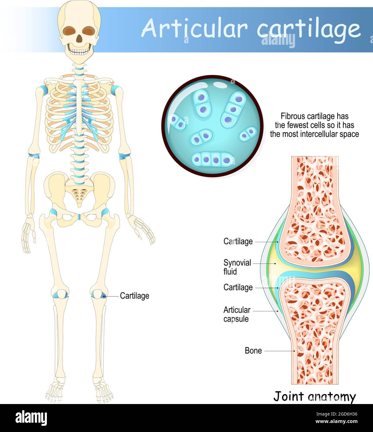
Cartilage. Human skeleton with articular cartilage. Joint anatomy

Schematic drawing of the locations of measurement of cartilage
A Fetal Skeleton Begins Entirely As Cartilage.
2.9K Views 4 Years Ago Vadnagar.
Web During Embryonic Development, Hyaline Cartilage Serves As Temporary Cartilage Models That Are Essential Precursors To The Formation Of Most Of The Axial And Appendicular Skeleton.
Web This Result Snaps A Four Match Losing Streak, And This Combined With Newcastle’s Draw Means They Only Need One Point From Their Last Two Games (At City, Home To Sheffield United) To Clinch Fifth.
Related Post: