Cervix Drawing
Cervix Drawing - The functions of these organs. Web the cervix is the lower portion (or the neck) of the uterus. It leads to the outside of the body. The cervix is approximately 4 cm in length and 3 cm in diameter. When the woman isn’t ovulating, the. Parts of the vagina are made of collagen and elastin, which help it expand during sexual stimulation and childbirth. It enables a baby to leave your uterus so that it can travel through your vagina (birth canal) during childbirth. Web the cervix is a fibromuscular organ that forms a canal between the lower, narrow end of the uterus and the vagina. The following 18 files are in this category, out of 18 total. During menstruation, the cervix opens slightly to allow menstrual blood to flow out of the uterus. Cervix during missed abortion.png 750 × 1,334; Web sometimes called the “neck of the uterus,” your cervix plays an important role in allowing fluids to pass between your uterus and vagina. Cervix diagram stock photos are available in a. When the woman isn’t ovulating, the. The female reproductive organs include several key structures, such as the ovaries, uterus, vagina, and. Web the cervix is the lower part of the uterus situated between the external os (external orifice) and internal os (internal orifice). The cervix is part of the female reproductive system. Web the cervix is a fibromuscular organ that forms a canal between the lower, narrow end of the uterus and the vagina. The endocervical canal (or endocervix) is the. Web sometimes called the “neck of the uterus,” your cervix plays an important role in allowing fluids to pass between your uterus and vagina. Anatomy of male and female urinary bladder, with labels. Similar to cervical cancer, cervical dysplasia is often related to hpv infection. Web cervical dysplasia is a condition in which abnormal cells grow on the surface of. Cervical effacement and dilation during delivery. Drawing of the anatomy of the cervix showing the internal os, endocervical canal, endocervix, ectocervix, and external os. Cervix changes from not effaced and dilated to fully effaced and totally cervix stock illustrations. There is also a pullout that shows a close up of the squamocolumnar junction (the area where the endocervix and ectocervix. The vagina is a muscular canal that connects the cervix and the uterus. The cervix is part of the female reproductive system. The cervical canal connects the interior of the vagina and the cavity of the body of uterus. The cervix plays vital roles in the control of movement into and out of the uterus, protection of the fetus. There. The functions of these organs. The uterus has a muscular outer layer called the myometrium and an inner lining called the endometrium. The organs in the female reproductive system include the uterus, ovaries, fallopian tubes, cervix, and vagina. There is also a pullout that shows a close up of the squamocolumnar junction (the area where the endocervix and ectocervix meet). It leads to the outside of the body. The female reproductive system is made up of the internal and external sex organs that function in reproduction of new offspring. Web sometimes called the “neck of the uterus,” your cervix plays an important role in allowing fluids to pass between your uterus and vagina. Web the cervix of the uterus is. The following 18 files are in this category, out of 18 total. The uterus and vagina are also shown. Web ovary, female reproductive system. Female reproductive system of internal organs continuous line drawing vector illustration isolated female reproductive system of internal organs continuous line. The opening in the ectocervix, the external os, marks the transition from the ectocervix to the. Cervix during missed abortion.png 750 × 1,334; The cervix is part of the female reproductive system. During menstruation, the cervix opens slightly to allow menstrual blood to flow out of the uterus. Web the ectocervix is the portion of the cervix that projects into the vagina. Web the cervix is the lower portion (or the neck) of the uterus. The female reproductive organs include several key structures, such as the ovaries, uterus, vagina, and vulva. The uterus and vagina are also shown. Female reproductive system of internal organs continuous line drawing vector illustration isolated female reproductive system of internal organs continuous line. Web the cervix is the lower portion (or the neck) of the uterus. Its name, cervix, comes. Web a quick, functional drawing of the cervical plexus of nerves in the neck. Although it is described as being cylindrical in shape, the anterior and posterior walls are more often ordinarily apposed. The female reproductive system is made up of the internal and external sex organs that function in reproduction of new offspring. The cervix is also a common site for cell changes that may indicate cancer. 3d illustartion of human femail reproductive system anatomy. Its name, cervix, comes from the latin word meaning “neck” due to its role as the narrow connection between the larger body of the uterus above the vagina below. There is also a pullout that shows a close up of the squamocolumnar junction (the area where the endocervix and ectocervix meet) and the cells that line the. Web squamous epithelial cells of human cervix under the microscope view. It is not cancer, but it is considered a precancerous condition. It leads to the outside of the body. The cervix is the lower part of the uterus that separates the lower uterus and the vagina. Drawing of the anatomy of the cervix showing the internal os, endocervical canal, endocervix, ectocervix, and external os. Female reproductive system of internal organs continuous line drawing vector illustration isolated female reproductive system of internal organs continuous line. The functions of these organs. Anatomy of male and female urinary bladder, with labels. The organs in the female reproductive system include the uterus, ovaries, fallopian tubes, cervix, and vagina.
Printable Cervix Diagrams HD 101 Diagrams

Cervical Ectropion Causes, Symptoms & Cervical Ectropion Treatment

Vector isolated illustration of female reproductive system anatomy

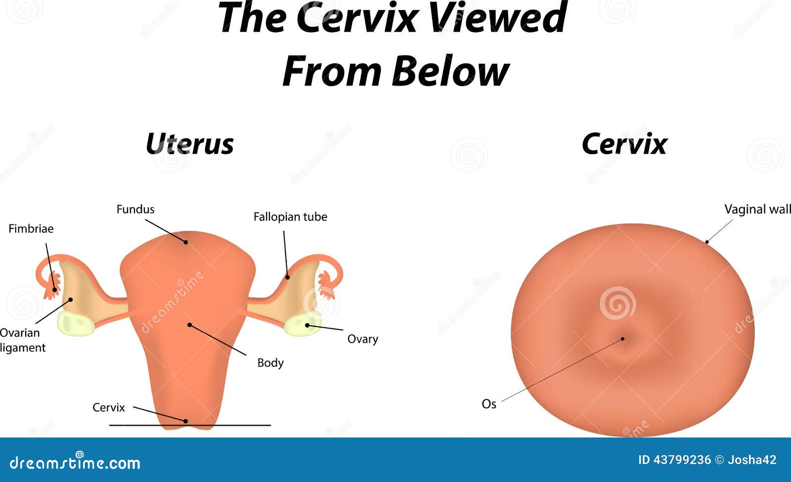
The Cervix stock vector. Illustration of internal, ovary 43799236
![[DIAGRAM] Cervix Diagram Side](https://intimateartscenter.com/wp-content/uploads/2016/04/Female-Internal-Organs-Side-View-Color_-labels2.jpg)
[DIAGRAM] Cervix Diagram Side

Image result for parts of cervix Anatomy, Human body anatomy, Body

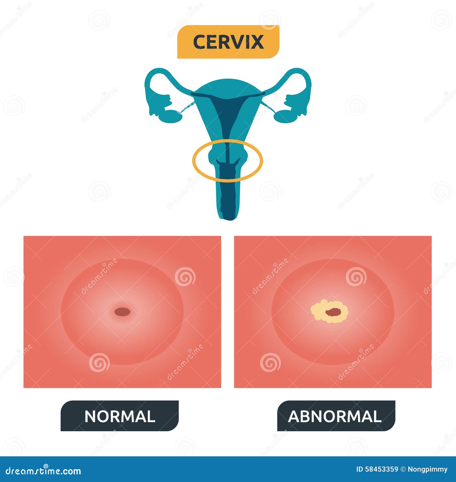
Cervix stock vector. Illustration of normal, smear, medical 58453359

Female Reproductive Tract (Anatomy) Gross Anatomy Flashcards ditki

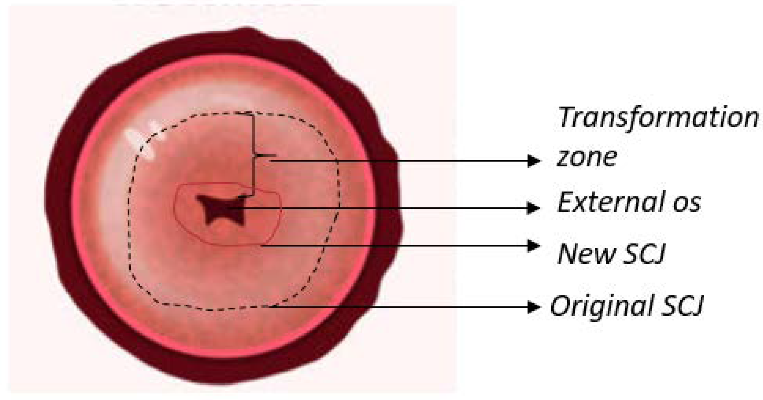
Cervical Imaging in the Low Resource Setting Encyclopedia MDPI

Cervix, Illustration Stock Photo Alamy
1 Cm, About The Size Of A Cheerio.
Web The Cervix Is A Fibromuscular Organ That Forms A Canal Between The Lower, Narrow End Of The Uterus And The Vagina.
Cervix Changes From Not Effaced And Dilated To Fully Effaced And Totally Cervix Stock Illustrations.
The Uterus Has A Muscular Outer Layer Called The Myometrium And An Inner Lining Called The Endometrium.
Related Post: