Drawing Of Stratified Squamous Epithelium
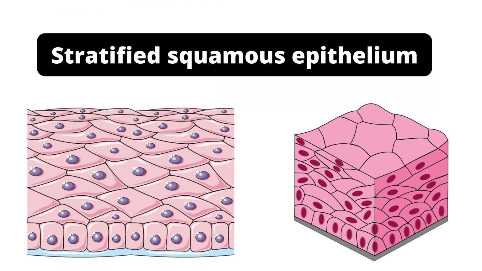
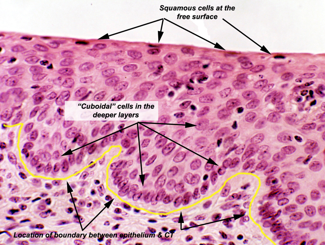
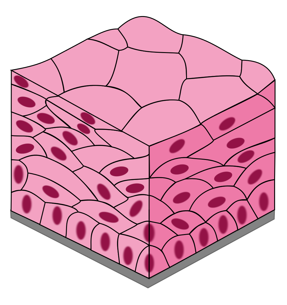
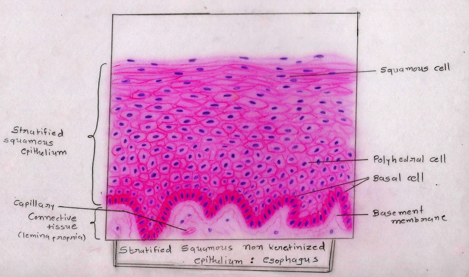
Drawing Of Stratified Squamous Epithelium - The stratified epithelium is named by the shape of the most apical layer of cells, closest to the free space. Web stratified squamous epithelium is part of two important protective membranes, where it exists in different forms: The apical cells appear squamous, whereas the basal layer contains either columnar or cuboidal cells. Web stratified squamous epithelium occurs on numerous surfaces where it provides an important protective function. Only one layer is in contact with the basement membrane; Anatomy with amrutha & joseph. Web stratified epithelia consist of multiple layers of cells, with one layer anchored to the basement membrane, known as the basal layer. A stratified epithelium consists of several stacked layers of cells. Keratin is a fibrous protein that provides strength and durability to our hair, skin, and nails. It is continuously replacing itself by division of the basal layer of cells. The stratified epithelium is named by the shape of the most apical layer of cells, closest to the free space. 3.8k views 3 years ago. Web the stratified squamous epithelium under a microscope shows the multiple layers of cells where the superficial one is flattened. Draw your structures proportionately to their size in your microscope’s field of view. Use the. Web if it is a stratified epithelium draw all the layers. 19k views 2 years ago cell biology. The stratified epithelium is named by the shape of the most apical layer of cells, closest to the free space. Only one layer is in contact with the basement membrane; Web stratified squamous epithelium occurs on numerous surfaces where it provides an. These cells change shaped as they move toward the surface and are eventually shed. You will also find the basal columnar cells in this stratified squamous epithelium. Epithelial cells are also classified by their shape: The top layer may be covered with dead cells containing keratin. In many cases, adjacent epithelial cells are linked by tight junctions so that the. The other layers adhere to one another to maintain structural integrity. Only one layer is in contact with the basement membrane; Its name arises from the squamous appearance of the outermost layer of cells. This type of epithelium comprises the epidermis of the skin. Underlying cell layers can be made of cuboidal or columnar cells as well. 19k views 2 years ago cell biology. Location and examples of stratified squamous epithelium. Web figure 5.4 epidermis the epidermis is epithelium composed of multiple layers of cells. These cells change shaped as they move toward the surface and are eventually shed. It is continuously replacing itself by division of the basal layer of cells. Web if it is a stratified epithelium draw all the layers. Squamous = top layer is flat. Web stratified squamous epithelium is the most common type of stratified epithelium in the human body. Skin that has four layers of cells is referred to as “thin skin.” It is made of four or five layers of epithelial cells, depending on its. This type of epithelium comprises the epidermis of the skin. The basal layer consists of cuboidal cells, whereas the outer layers are squamous, keratinized cells, so the whole epithelium is often described as. Anatomy with amrutha & joseph. Download the complete guide to neet ug prep. This epithelium contains 5 layers: Location and examples of stratified squamous epithelium. In many cases, adjacent epithelial cells are linked by tight junctions so that the epithelium forms a barrier that regulates the movement or substances. Underlying cell layers can be made of cuboidal or columnar cells as well. The keratinization, or lack thereof, of the apical surface domains of the cells. Web the stratified. The keratinization, or lack thereof, of the apical surface domains of the cells. Fill in the blanks next to your drawing. Download the complete guide to neet ug prep. A typical example of stratified squamous keratinized epithelium is the epidermis. Anatomy with amrutha & joseph. Web stratified squamous epithelium is the most common type of stratified epithelium in the human body. These cells change shaped as they move toward the surface and are eventually shed. This epithelium protects against physical and chemical wear and tear. It is made of four or five layers of epithelial cells, depending on its location in the body. Stratified squamous. Web figure 5.4 epidermis the epidermis is epithelium composed of multiple layers of cells. Web stratified squamous nonkeratinized epithelium. Download the complete guide to neet ug prep. The top layer may be covered with dead cells containing keratin. The keratinization, or lack thereof, of the apical surface domains of the cells. Location and examples of stratified squamous epithelium. The basal layer consists of cuboidal cells, whereas the outer layers are squamous, keratinized cells, so the whole epithelium is often described as. 3.8k views 3 years ago. Draw your structures proportionately to their size in your microscope’s field of view. Stratified squamous epithelium is the most common type of stratified. In many cases, adjacent epithelial cells are linked by tight junctions so that the epithelium forms a barrier that regulates the movement or substances. Stratified squamous epithelia are tissues formed from multiple layers of cells resting on a basement membrane, with the superficial layer (s) consisting of squamous cells. Use the image slider below to learn how to use a microscope to identify and study nonkeratinized stratified squamous epithelium lining the esophagus. This epithelium contains 5 layers: The other layers adhere to one another to maintain structural integrity. Web if it is a stratified epithelium draw all the layers.
Illustration of Stratified Squamous Epithelium Stock Photo Alamy

How to draw stratified squamous epithelium easy way YouTube
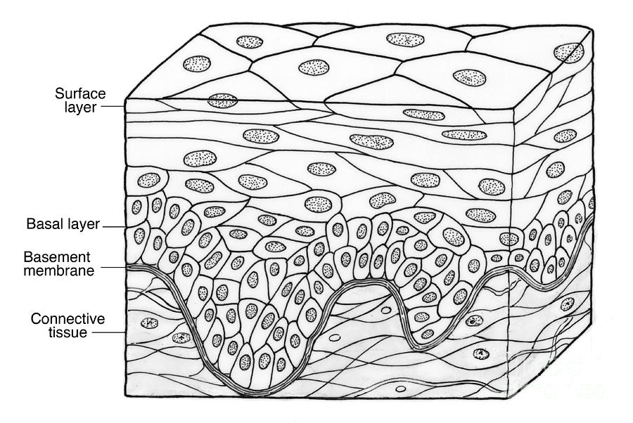
Illustration Of Stratified Squamous Photograph by Science Source Pixels

Stratified squamous epithelium, illustration Stock Image F024/3098

Oral mucosa Lamina propria Wikipedia Basement membrane
Stratified squamous epithelium Function, Definition, Location, Types.
Epithelial Tissue Locations Stratified Squamous Epithelium Histology
Epithelial tissues

Stratified Squamous Epithelium Overview, Function & Location Lesson
Histology Image Membranous epithelium
What Are Squamous Epithelial Cells?
A Typical Example Of Stratified Squamous Keratinized Epithelium Is The Epidermis.
Web Stratified Squamous Epithelium Is Part Of Two Important Protective Membranes, Where It Exists In Different Forms:
You Will Also Find The Basal Columnar Cells In This Stratified Squamous Epithelium.
Related Post: