Drawing Of The Pancreas
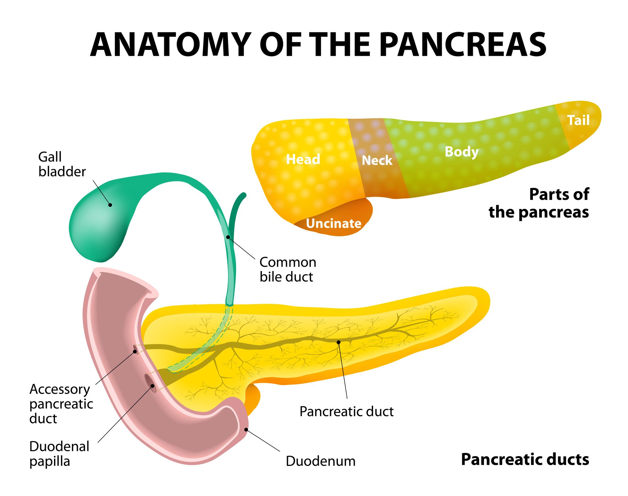
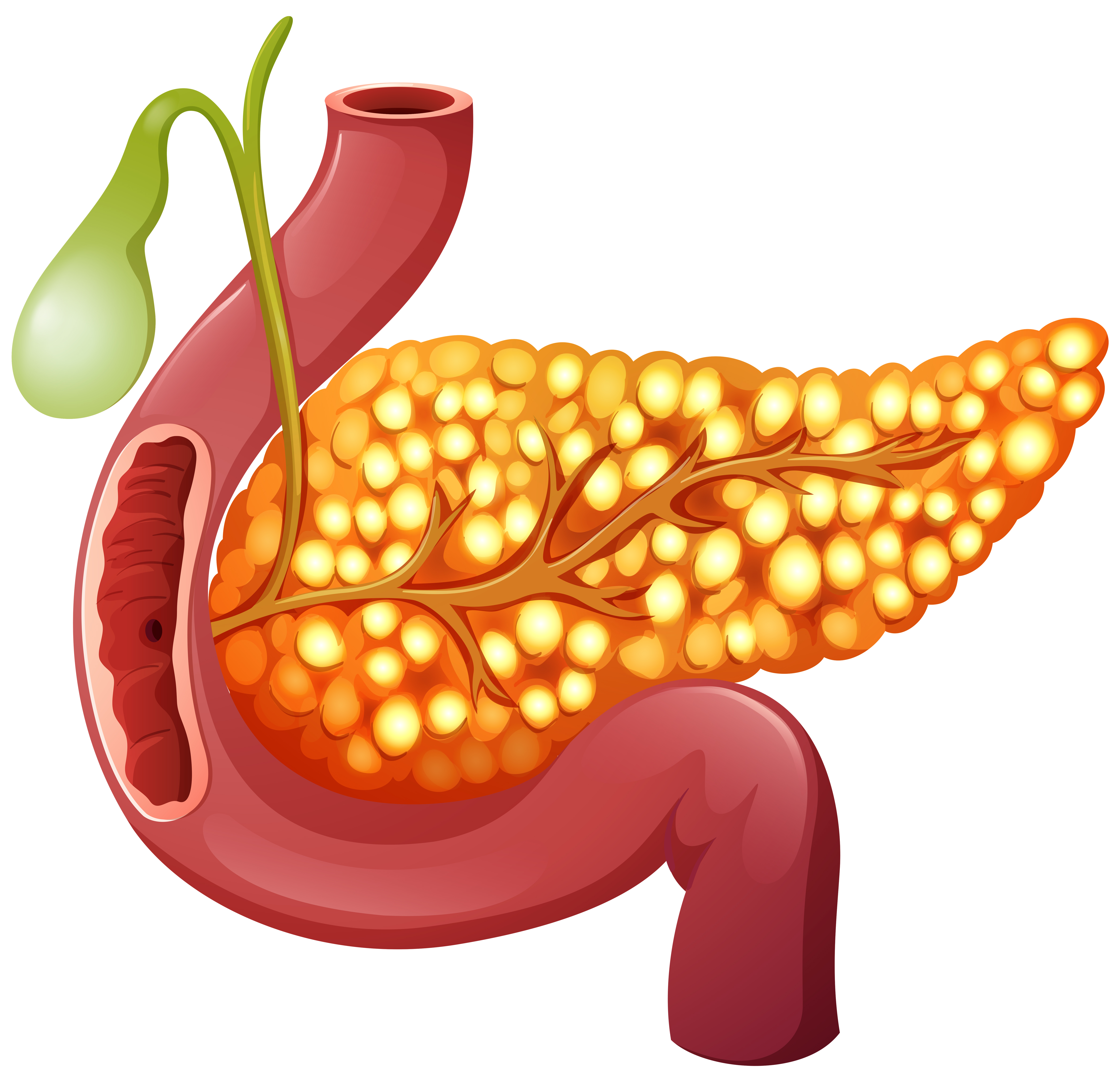
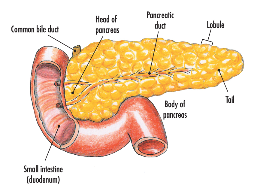
Drawing Of The Pancreas - The exocrine pancreas is a complex tubular network. It sits within the curve of the duodenum (the first part of the small intestine) and is divided into two parts: The head, uncinate process, neck, body and tail. The pancreas is a large, mixed gland composed of five parts: Web the pancreas is a long, slender organ, most of which is located posterior to the bottom half of the stomach ( figure 17.9.1 ). The pancreas is supplied by pancreatic arteries stemming from surrounding vessels and is innervated by the vagus. Histology (also known as microscopic anatomy or microanatomy) is the branch of biology that studies the microscopic anatomy of biological tissues; Within the abdomen, the pancreas has direct anatomical relations to several. Exocrine glandular tissues in the pancreas produce pancreatic enzymes that are dumped into the small intestine via the pancreatic duct. The parotid gland is a vital exocrine organ and the primary site for salivary gland tumors. “alcohol causes damage to the tissues over time which can lead to changes in the cell's dna and increased risk for cancer,” tatum says. It is composed of several parts. The pancreas is located below and behind the stomach. Web the pancreas is a gland that lies across the back side of the abdomen. Histology (also known as microscopic anatomy. Web the pancreas is a long, slender organ, most of which is located posterior to the bottom half of the stomach ( figure 17.9.1 ). Although it is primarily an exocrine gland, secreting a variety of digestive enzymes, the pancreas also has endocrine cells. This drawing depicts a configuration that is intermediate to those shown in fig. Schematic drawings of. The blackboard has a slight texture. Within the abdomen, the pancreas has direct anatomical relations to several. The head, body, and tail. Exocrine glandular tissues in the pancreas produce pancreatic enzymes that are dumped into the small intestine via the pancreatic duct. Its pancreatic islets —clusters of cells formerly known as the islets of langerhans. The head proper and the uncinate. Pancreas drawing stock photos are available in a variety of sizes and formats to fit your needs. The enzymes are necessary to digest food. Although it is primarily an exocrine gland, secreting a variety of digestive enzymes, the pancreas has an endocrine function. Human internal organs, tooth and eye. Web although the regions are not labeled we see the head of the pancreas at left, and tail of the pancreas, image image right. It forms an integral part of the digestive system. Growth and rotations (arrows) of the duodenum bring the ventral pancreatic bud toward the dorsal. The neck is the constricted part between the. The pancreas is a. The pancreas is a large, mixed gland composed of five parts: The pancreas is a retroperitoneal organ and does not have a capsule. Although it is primarily an exocrine gland, secreting a variety of digestive enzymes, the pancreas also has endocrine cells. To investigate the molecular characteristics of and potential for precision medicine in kras wildtype pancreatic ductal adenocarcinoma (pdac).. Histology (also known as microscopic anatomy or microanatomy) is the branch of biology that studies the microscopic anatomy of biological tissues; Web antique illustration of human body anatomy: The endocrine portion is arranged as discrete islets of langerhans, which are composed of five different endocrine cell types (alpha, beta, delta, epsilon, and upsilon) secreting at least five hormones including glucagon,. The head, body, and tail. It forms an integral part of the digestive system. Web the pancreas is a glandular organ that produces a number of hormones essential to the body. The illustration in figure 1 demonstrates the anatomical relationships between the pancreas and organs surrounding it in the abdomen. Endocrine function of the pancreas involves the. Web the pancreas is an elongated, tapered organ located across the back of the belly, behind the stomach. Clinical data were obtained via chart review. The pancreas is an elongated gland located deep within the abdomen, tucked in between the stomach and the spine. Pancreas drawing stock photos are available in a variety of sizes and formats to fit your. It lies posterior to the pyloric part of the stomach. Web #pancreas #howtodraw #adimushowthis is an easy drawing of pancreas. Web the histology of the pancreas. Web the pancreas is a glandular organ that produces a number of hormones essential to the body. Web anatomy of the pancreas. The parotid gland is a vital exocrine organ and the primary site for salivary gland tumors. Web the pancreas is a composite organ, which has exocrine and endocrine functions. The bile duct and pancreatic duct are also shown. The second and third portions of the duodenum curve around the head of the pancreas. The pancreas lies in the epigastrium or upper central region of the abdomen. The head, body, and tail. One end of the pancreas is wider than the other and is called the head: This drawing depicts a configuration that is intermediate to those shown in fig. Postpancreatectomy hemorrhage (pph) following the development of postoperative pancreatic fistula is a major complication, often. The exocrine pancreas is a complex tubular network. Histology (also known as microscopic anatomy or microanatomy) is the branch of biology that studies the microscopic anatomy of biological tissues; The blackboard has a slight texture. This royalty free vector illustration features the human body vector icon set on black chalkboard. The right side of the organ—called the head—is the widest part of the organ and lies in the curve of the duodenum, the first division of the small intestine. The pancreas is supplied by pancreatic arteries stemming from surrounding vessels and is innervated by the vagus. This short section that lies between the head and the body is around 2.5 cm long.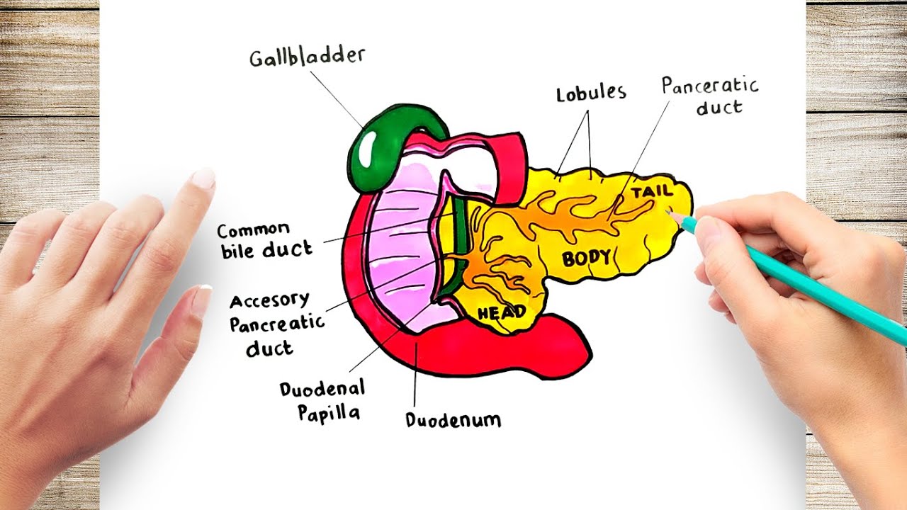
How To Draw A Pancreas YouTube
Pictures Of The Pancreas Beating Pancreatitis

Pancreas anatomical cross section model, vector illustration medical
A healthy human Pancreas 303570 Vector Art at Vecteezy
/pancreas_lg-595e8eae3df78c4eb64f2fce.jpg)
Pancreas Anatomy and Function

Pancreas Vector Image Healthcare Illustrations Creative Market

The Pancreas Anatomy Anatomy Book

Human Pancreas drawing How to draw human Pancreas Human pancreas

Pancreas Medical anatomy, Human anatomy and physiology, Anatomy
The pancreas Anatomy of the pancreas Structure of the pancreas
Schematic Drawings Of The Successive Stages In The Development Of The Pancreas From The Fifth Through The Eighth Weeks.
Diagrammatic Transverse Sections Through The Duodenum And The Developing Pancreas.
The Endocrine Portion Is Arranged As Discrete Islets Of Langerhans, Which Are Composed Of Five Different Endocrine Cell Types (Alpha, Beta, Delta, Epsilon, And Upsilon) Secreting At Least Five Hormones Including Glucagon, Insulin, Somatostatin, Ghrelin, And.
The Pancreas Is Located Below And Behind The Stomach.
Related Post: