Embryology Drawing
Embryology Drawing - For example, the heart begins to beat in week 4, the eyes begin to form in week 5, and the lungs begin to form in week 6. Once you have a solid roadmap of development based on the highlights, the details will. A zygote is the single cell formed when an egg and a sperm cell fuse; Web mesoderm from medial to lateral: Web embryo drawing is the illustration of embryos in their developmental sequence. • sclerotome derives the bone of the axial skeleton: As we discover in haeckel’s embryos, german biologist ernst haeckel included illustrations of the embryological stages of vertebrates in a series of books published between 1868. Web click here to listen to the full podcast. Embryos and the development of embryos of various species within a class are similar even if their. Embryo images normal and abnormal mammalian development is a tutorial that uses scanning electron micrographs (sems) as the primary resource to teach mammalian embryology. Darwin used the science of embryology to support his conclusions. As we heard in episode 27 of our last series, his tree of life diagram was widely reproduced, despite not representing darwinian principles. Web accuracy in embryo illustrations. Web in embryology, some highlights are particularly easy to identify, because each week has milestone events that are frequently tested. Embryos and. Embryo images normal and abnormal mammalian development is a tutorial that uses scanning electron micrographs (sems) as the primary resource to teach mammalian embryology. Web embryo drawing is the illustration of embryos in their developmental sequence. Web embryo images normal and abnormal mammalian development is a tutorial that uses scanning electron micrographs (sems) as the primary resource to teach mammalian. Web embryo drawing is the illustration of embryos in their developmental sequence. In animals, the zygote divides repeatedly to form a ball of cells, which then forms a set of tissue layers that migrate and fold to form an early. As we discover in haeckel’s embryos, german biologist ernst haeckel included illustrations of the embryological stages of vertebrates in a. Web in haeckel’s embryos: Web in embryology, some highlights are particularly easy to identify, because each week has milestone events that are frequently tested. An embryo at the end of 7 weeks of development is only 10 mm in length, but its developing eyes, limb buds, and tail are already visible. The zygote’s first priority is dividing to make lots. Web 8.2.1 in the nineteenth century. Web mesoderm from medial to lateral: Soemmerring points out the presence of a thick umbilical cord, which may relate to the structures containing the cord in the 5th week pf, such as umbilical vessels, allantois, vitelline duct, and vitelline vessels. He recaptures the shocking novelty of pictures that enthralled. 3d models, histology sections and. Web in haeckel’s embryos: An embryo at the end of 7 weeks of development is only 10 mm in length, but its developing eyes, limb buds, and tail are already visible. Web embryology is the study and analysis of embryos. He tracks the drawings and the charges against them from their genesis in the 19th century to the present day.. Ditki is the ideal resource for the flipped classroom: It has been widely noted that a number of the embryos in top row of the tables 6 and 7 from haeckel's anthropogenie (1874) are not realistic representations. Embryos and the development of embryos of various species within a class are similar even if their. This period is also considered the. Evidence of an evolutionary common ancestor is seen in the similarity of embryos in markedly different species. Web by the end of the embryonic period, the embryo is approximately 3 cm (1.2 in) from crown to rump and weighs approximately 8 g (0.25 oz). Web in embryology, some highlights are particularly easy to identify, because each week has milestone events. Web embryo images normal and abnormal mammalian development is a tutorial that uses scanning electron micrographs (sems) as the primary resource to teach mammalian embryology. Web mesoderm from medial to lateral: Web embryo drawing is the illustration of embryos in their developmental sequence. The somites (the paraxial mesoderm) form the axial musculoskeleton and dermis as follows: Ditki is the ideal. He tracks the drawings and the charges against them from their genesis in the 19th century to the present day. Embryo drawing is the illustration of embryos in their developmental sequence.in plants and animals, an embryo develops from a zygote, the single cell that results when an egg and sperm fuse during fertilization.in animals, the zygote divides repeatedly to form. As we discover in haeckel’s embryos, german biologist ernst haeckel included illustrations of the embryological stages of vertebrates in a series of books published between 1868. 3d models, histology sections and gene expression data in human embryonic and fetal development. Web in embryology, some highlights are particularly easy to identify, because each week has milestone events that are frequently tested. It has been widely noted that a number of the embryos in top row of the tables 6 and 7 from haeckel's anthropogenie (1874) are not realistic representations. Web mesoderm from medial to lateral: This period is also considered the organogenic period, when most organs within the embryo have begun to form. Fast shippingdeals of the dayread ratings & reviewsshop best sellers • sclerotome derives the bone of the axial skeleton: However, the assertion by explore evolution that haeckel claimed that top row represented earliest embryos is false. In animals, the zygote divides repeatedly to form a ball of cells, which then forms a set of tissue layers that migrate and fold to form an early. For example, the heart begins to beat in week 4, the eyes begin to form in week 5, and the lungs begin to form in week 6. Soemmerring points out the presence of a thick umbilical cord, which may relate to the structures containing the cord in the 5th week pf, such as umbilical vessels, allantois, vitelline duct, and vitelline vessels. Web embryology is the study and analysis of embryos. Web embryo images normal and abnormal mammalian development is a tutorial that uses scanning electron micrographs (sems) as the primary resource to teach mammalian embryology. There are links to more detailed descriptions which can be viewed in a week. He tracks the drawings and the charges against them from their genesis in the 19th century to the present day.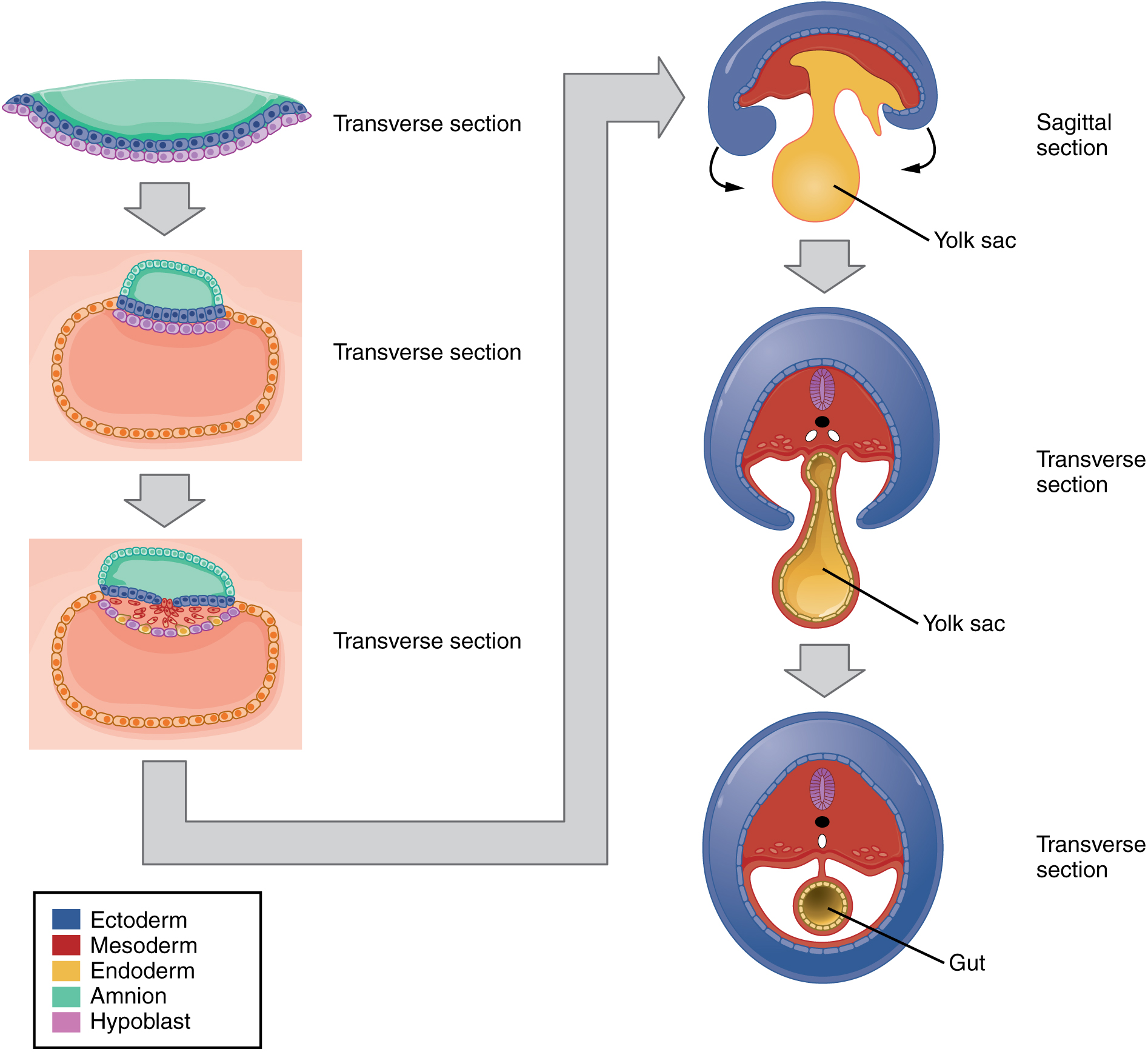
Embryonic Development · Anatomy and Physiology
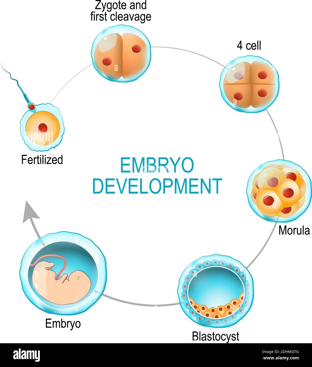
embryo development. from fertilization to zygote, morula and Blastocyst
Embryology Evolution

Stages in human embryonic development Royalty Free Vector
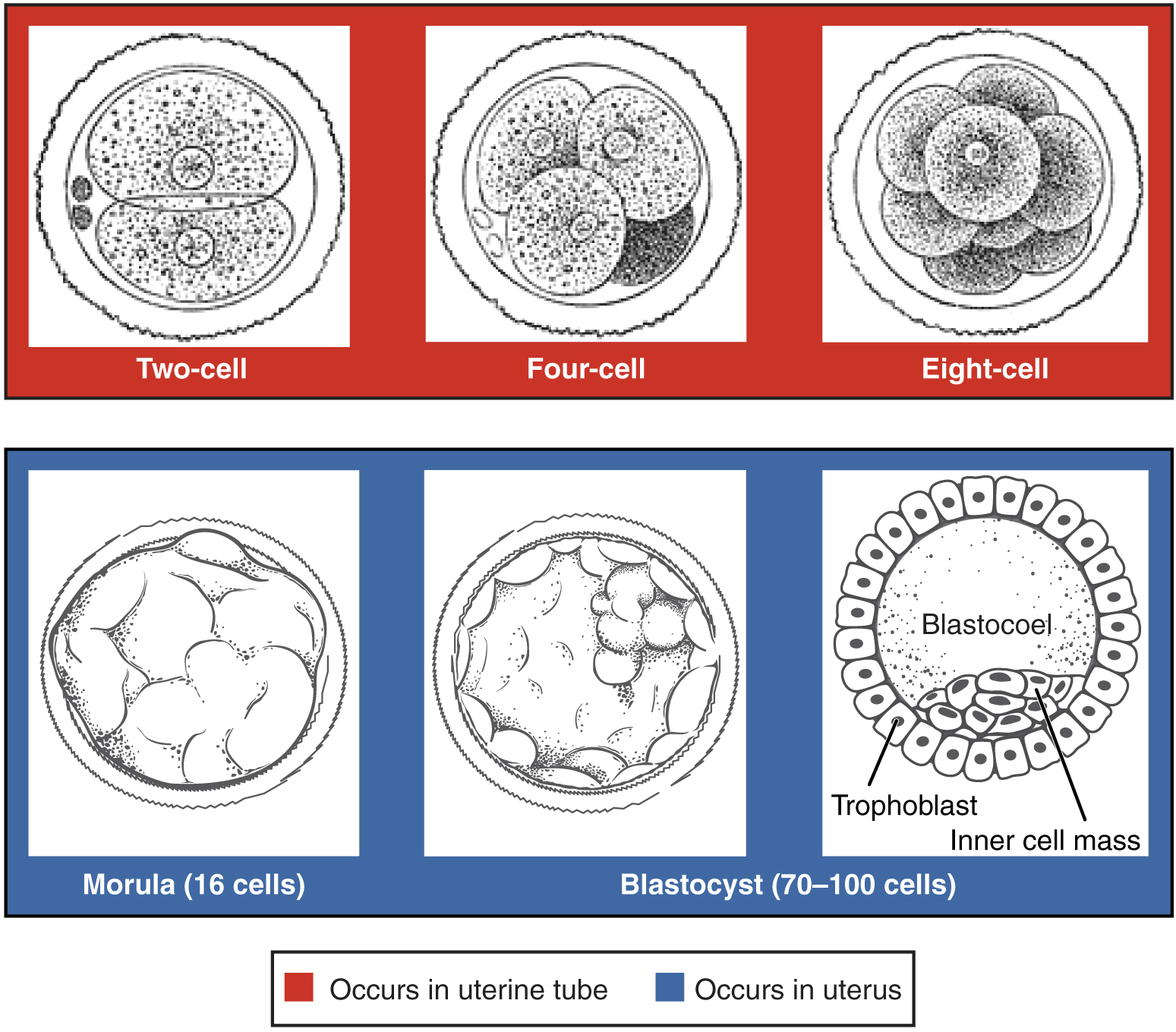
Embryonic Development · Anatomy and Physiology

Human embryo Royalty Free Vector Image VectorStock
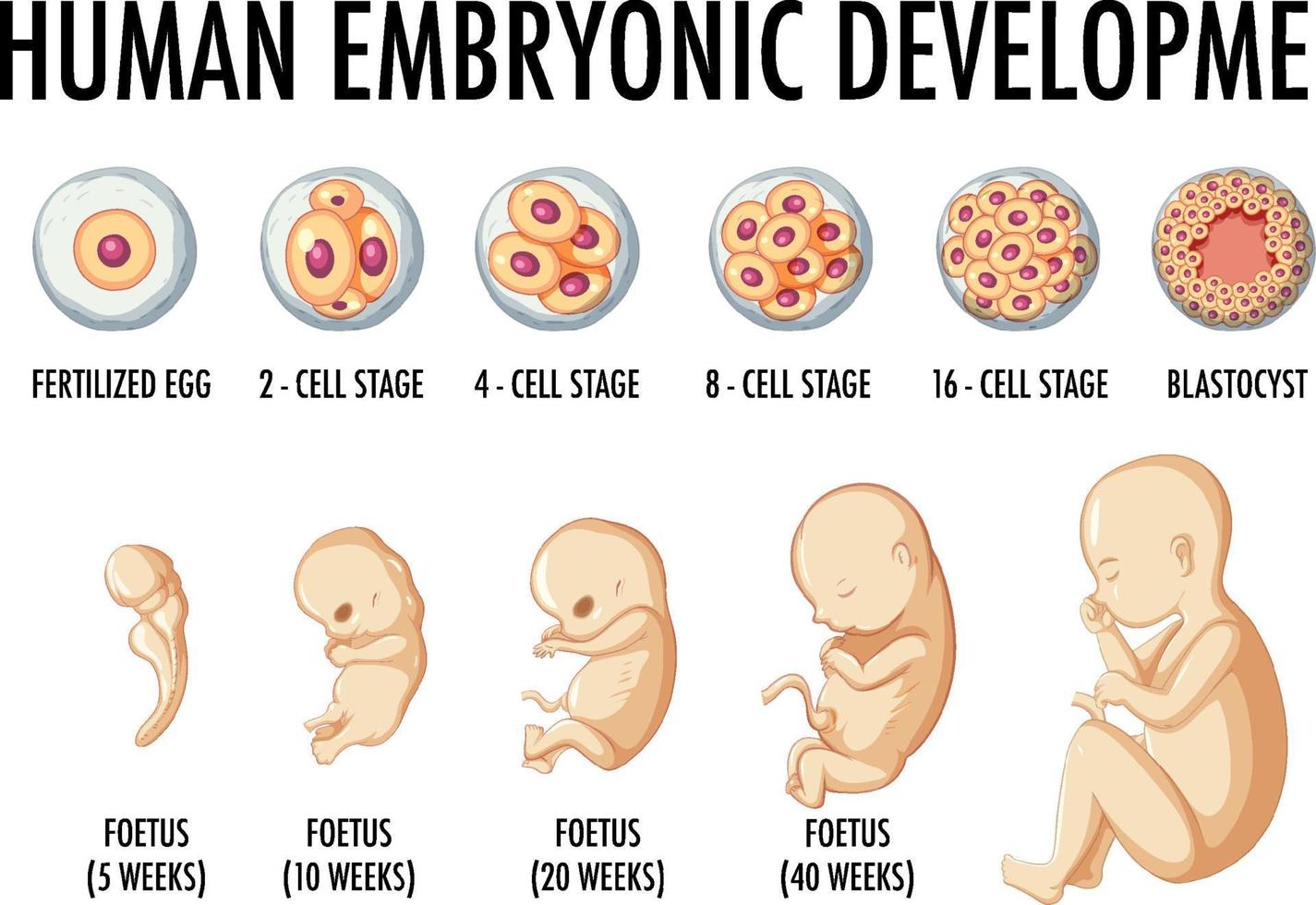
Human embryonic development in human infographic 6158571 Vector Art at
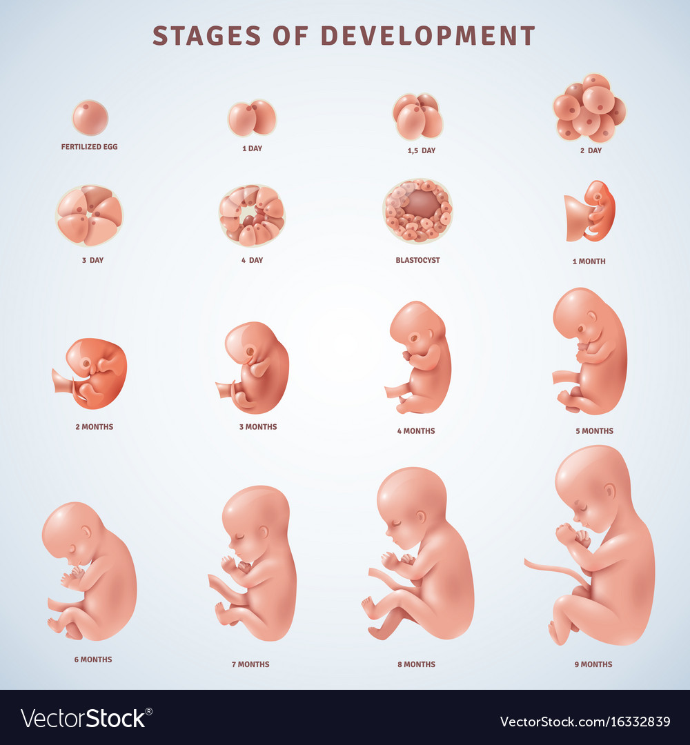
Stages human embryonic development Royalty Free Vector Image

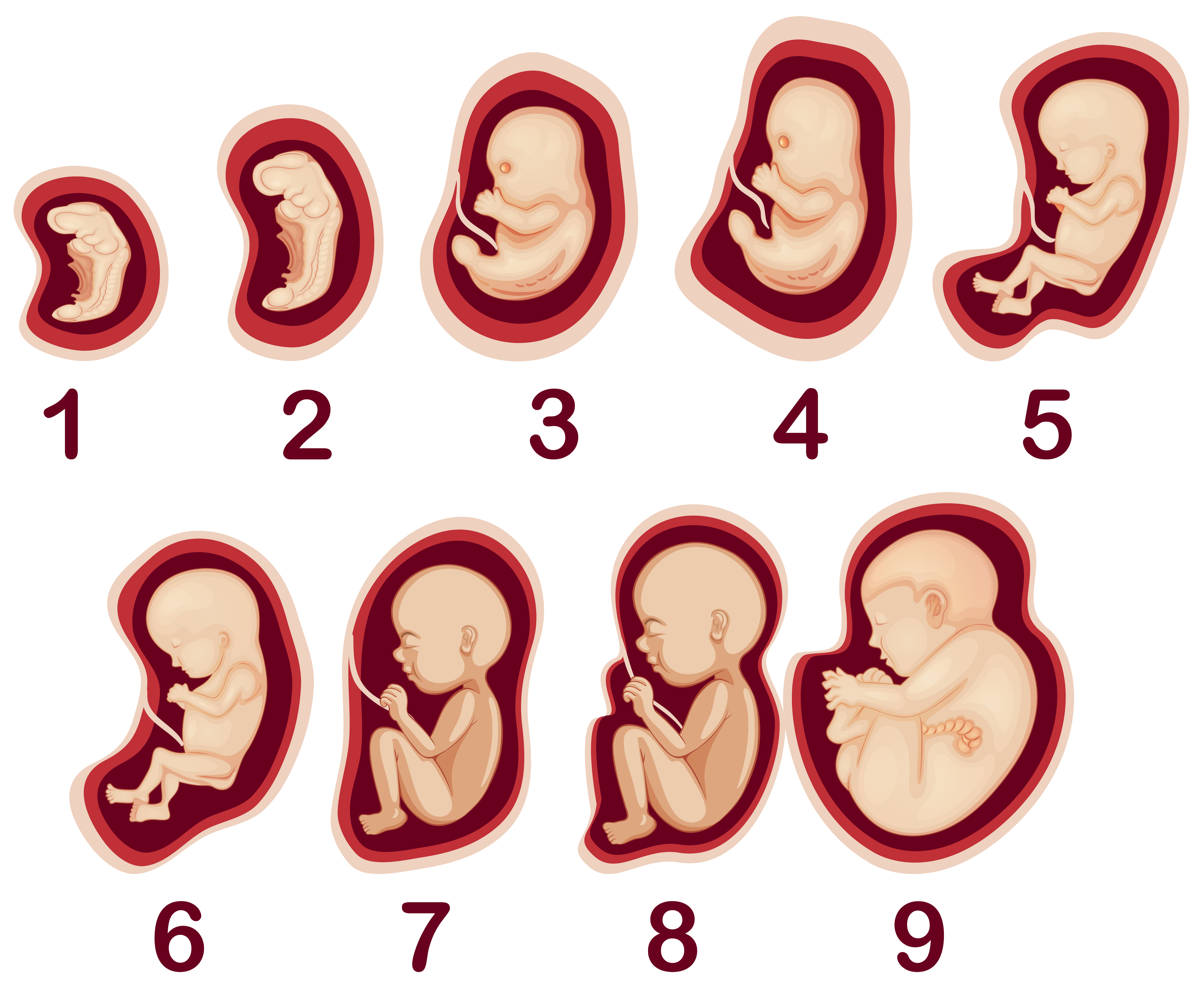
Embryo Development A Development process of Fetus Week by Week

Paper Description of a 4 mm human embryo (1906) Embryology
Images, Evolution And Fraud, Published By The University Of Chicago Press, Dr Nick Hopwood Tells The Full Story For The First Time.
Embryological Research Began In The Decades Around The Turn Of The Nineteenth Century.
The Zygote’s First Priority Is Dividing To Make Lots Of New Cells, So Its First Few Days Are Spent In Rapid Mitotic.
The Spine And The Posterior Base Of The Skull.
Related Post: