How To Draw A Prokaryotic Cell
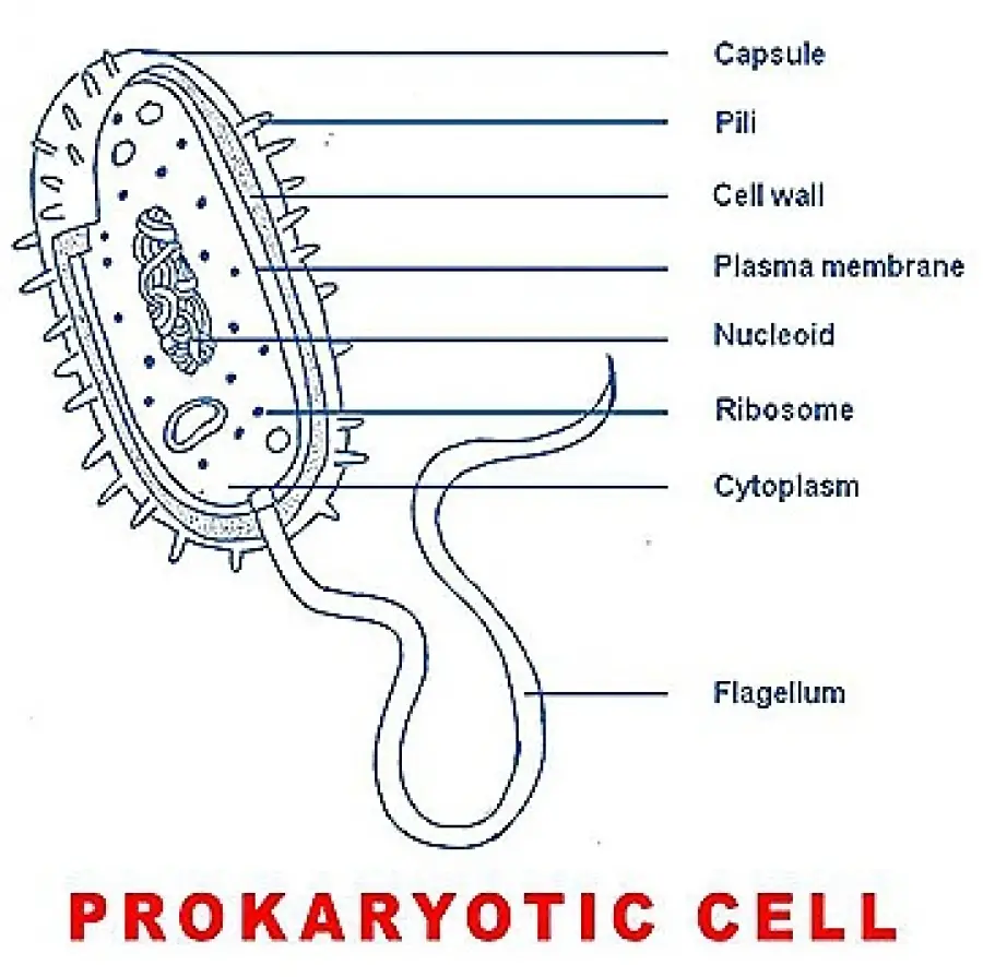
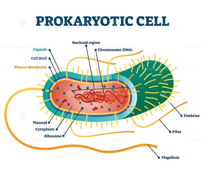
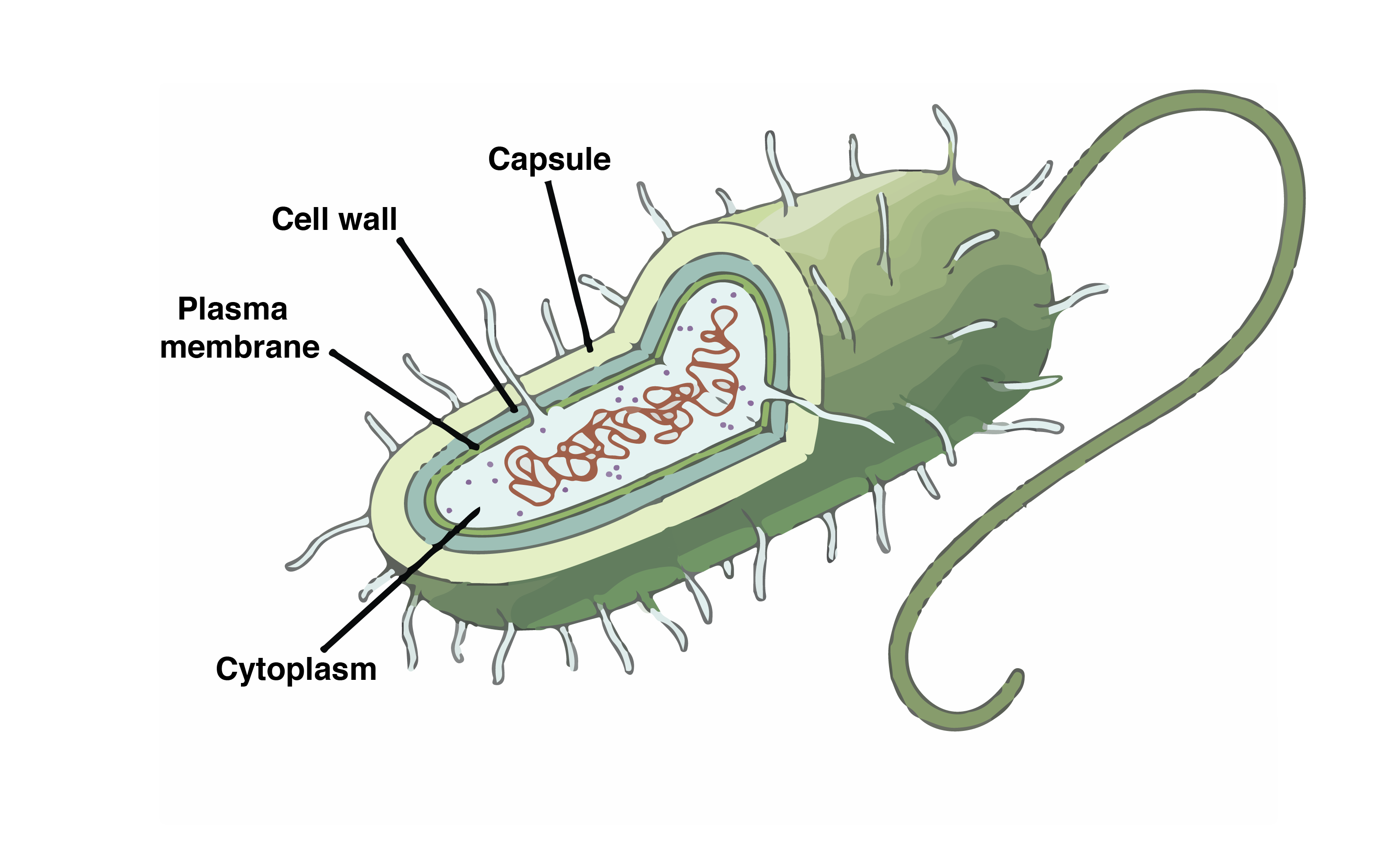
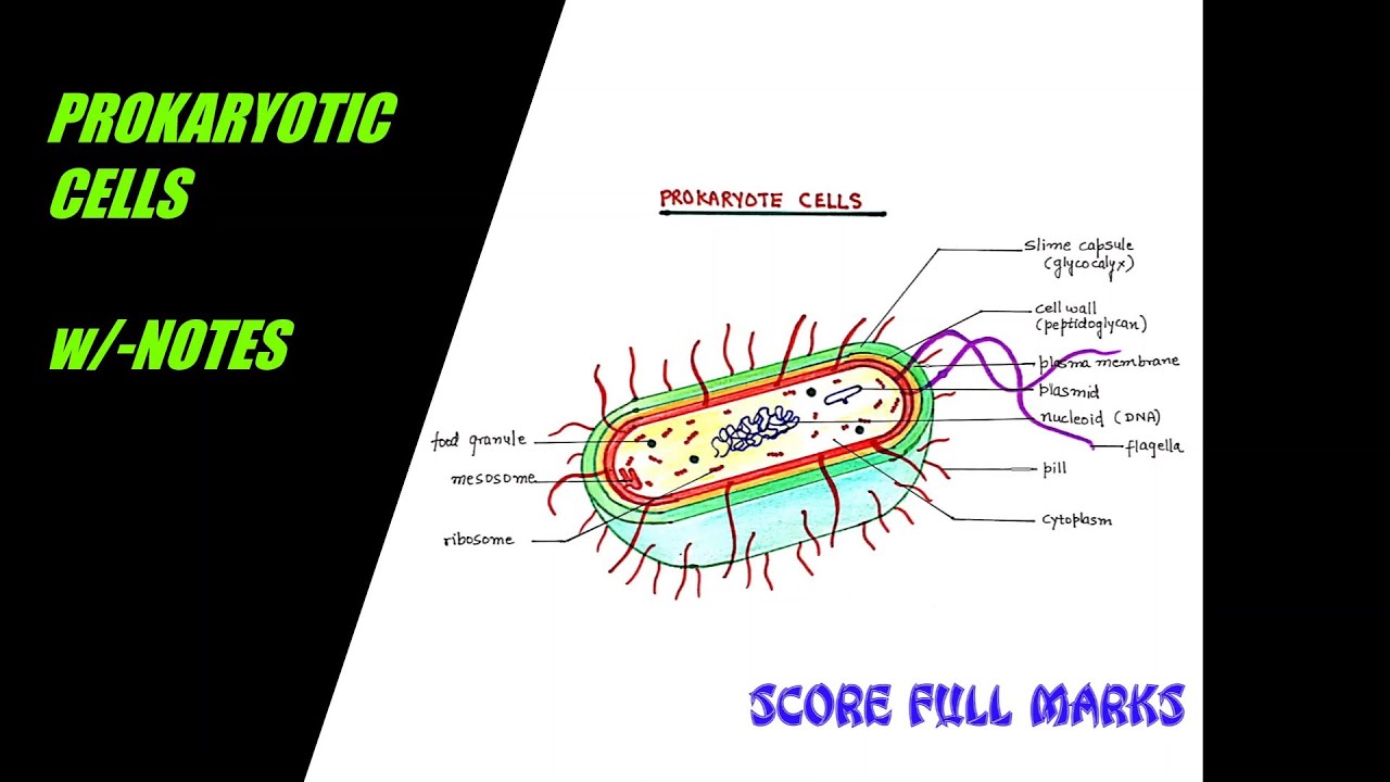
How To Draw A Prokaryotic Cell - The prokaryotic cell is a smaller and less complex type of cell which is associated with bacteria. Like other prokaryotic cells, this bacterial cell lacks a nucleus but has other cell parts, including a plasma membrane, cytoplasm, ribosomes, and dna. Read lab 4 and be ready to begin the lab exercise. Web how to draw a prokaryotic cell. How to draw prokaryotic cell / step by step drawing for beginners. Web prokaryotic dna is found in a central part of the cell: In your lab notebook diagram what you will do in lab 4. The three most common prokaryotic cell shapes are shown here. Prokaryotes fall into three basic categories based on their shape, visualized here using scanning electron microscopy: This diagram shows the structure of a typical prokaryotic cell, a bacterium. Prokaryotes fall into three basic categories based on their shape, visualized here using scanning electron microscopy: Web demonstrate how to draw cell structures seen with a microscope using sharp, carefully joined lines and straight edge lines for labels. Step by step and simple way to draw prokaryotic cell with easy methods.reference; Flow chart, diagram, series of cartoons, list, outline, etc.. Web how to draw prokaryotic cell step by step for beginners ! Prokaryotes fall into three basic categories based on their shape, visualized here using scanning electron microscopy: The most common shapes are helices, spheres, and rods (see figure below ). Web prokaryotic cells 2.2.1 draw and label a diagram of the ultrastructure of escherichia coli (e. Web typical prokaryotic. Like other prokaryotic cells, this bacterial cell lacks a nucleus but has other cell parts, including a plasma membrane, cytoplasm, ribosomes, and dna. Web how to draw prokaryotic cell / step by step drawing for beginners. These neat, well labelled and colorful diagrams will make your answers look more. 36k views 3 years ago class 9 diagram. All cells, which. Hello friends!!!!in this video, i will be showing you that how to draw a prokaryotic. Understand the primary differences between prokaryotic and eukaryotic cells. 1.7k views 3 years ago simple drawings. Web i am demonstrating the colorful diagram of prokaryotic cells step by step which you can draw very easily. The photosynthetic prokaryotes include cyanobacteria that perform photosynthesis. Some archaeal membranes are monolayer rather than bilayer. 12k views 1 year ago easy biology diagrams. This can take many different forms: Web about press copyright contact us creators advertise developers terms privacy policy & safety how youtube works test new features nfl sunday ticket press copyright. 36k views 3 years ago class 9 diagram. The photosynthetic prokaryotes include cyanobacteria that perform photosynthesis. (a) cocci, or spherical (a pair is shown); 36k views 3 years ago class 9 diagram. How to make a prokaryotic cell model. How to draw prokaryotic cell / step by step drawing for beginners. Prokaryotes fall into three basic categories based on their shape, visualized here using scanning electron microscopy: Web about press copyright contact us creators advertise developers terms privacy policy & safety how youtube works test new features nfl sunday ticket press copyright. (a) cocci, or spherical (a pair is shown); Step by step and simple way to draw prokaryotic cell with. Prokaryotes fall into three basic categories based on their shape, visualized here using scanning electron microscopy: This figure shows the generalized structure of a prokaryotic cell. What is a prokaryotic cell? Organisms within the domains bacteria and archaea are based on the prokaryotic cell, while all other forms of life are eukaryotic. 36k views 3 years ago class 9 diagram. Web how to draw prokaryotic cell / step by step drawing for beginners. 2.2.2 annotate the diagram from 2.2.1 with the functions of each named structure. Identify each of these parts in the diagram. Prokaryotes include bacteria and archaea. Step by step and simple way to draw prokaryotic cell with easy methods.reference; The figure below shows the sizes of prokaryotic, bacterial, and eukaryotic, plant and animal, cells as well as other molecules and organisms on a logarithmic. 12k views 1 year ago easy biology diagrams. Web typical prokaryotic cells range from 0.1 to 5.0 micrometers (μm) in diameter and are significantly smaller than eukaryotic cells, which usually have diameters ranging from 10. Prokaryotes fall into three basic categories based on their shape, visualized here using scanning electron microscopy: Like other prokaryotic cells, this bacterial cell lacks a nucleus but has other cell parts, including a plasma membrane, cytoplasm, ribosomes, and dna. Coli) as an example of a prokaryote. 1.7k views 3 years ago simple drawings. This diagram shows the structure of a typical prokaryotic cell, a bacterium. How to make a prokaryotic cell model. (a) cocci, or spherical (a pair is shown); Measure the field of view diameter of a microscope under low power. Web demonstrate how to draw cell structures seen with a microscope using sharp, carefully joined lines and straight edge lines for labels. Flow chart, diagram, series of cartoons, list, outline, etc. 36k views 3 years ago class 9 diagram. Web about press copyright contact us creators advertise developers terms privacy policy & safety how youtube works test new features nfl sunday ticket press copyright. This figure shows the generalized structure of a prokaryotic cell. Prokaryotes include bacteria and archaea. These neat, well labelled and colorful diagrams will make your answers look more. Read lab 4 and be ready to begin the lab exercise.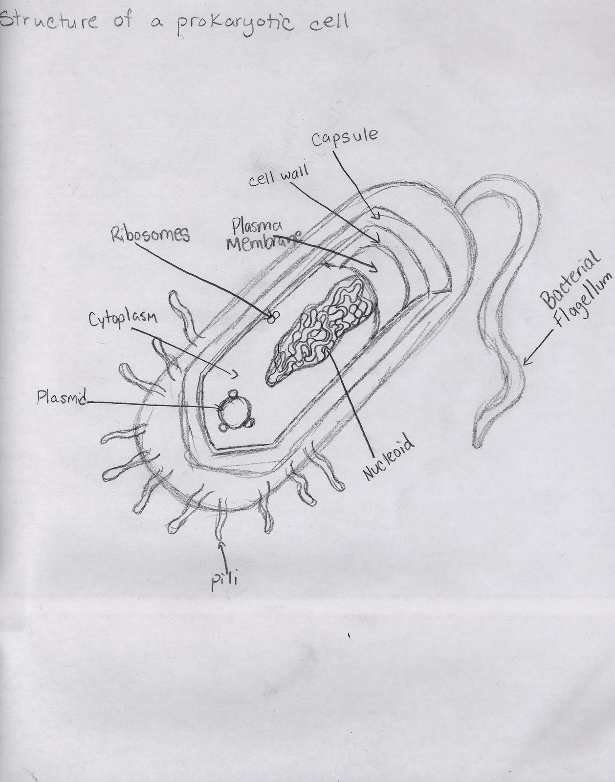
Cell Types and Structure Structure of Prokaryotic Cell
Simple Prokaryotic Cell Diagram
Prokaryotic cell structure diagram, vector illustration cross section

prokaryotic Cell diagram easy How to draw Prokaryotic Cell diagram

Prokaryotic Cell Diagram With Labels General Wiring Diagram
Prokaryotic Gene Structure Chloe's Science

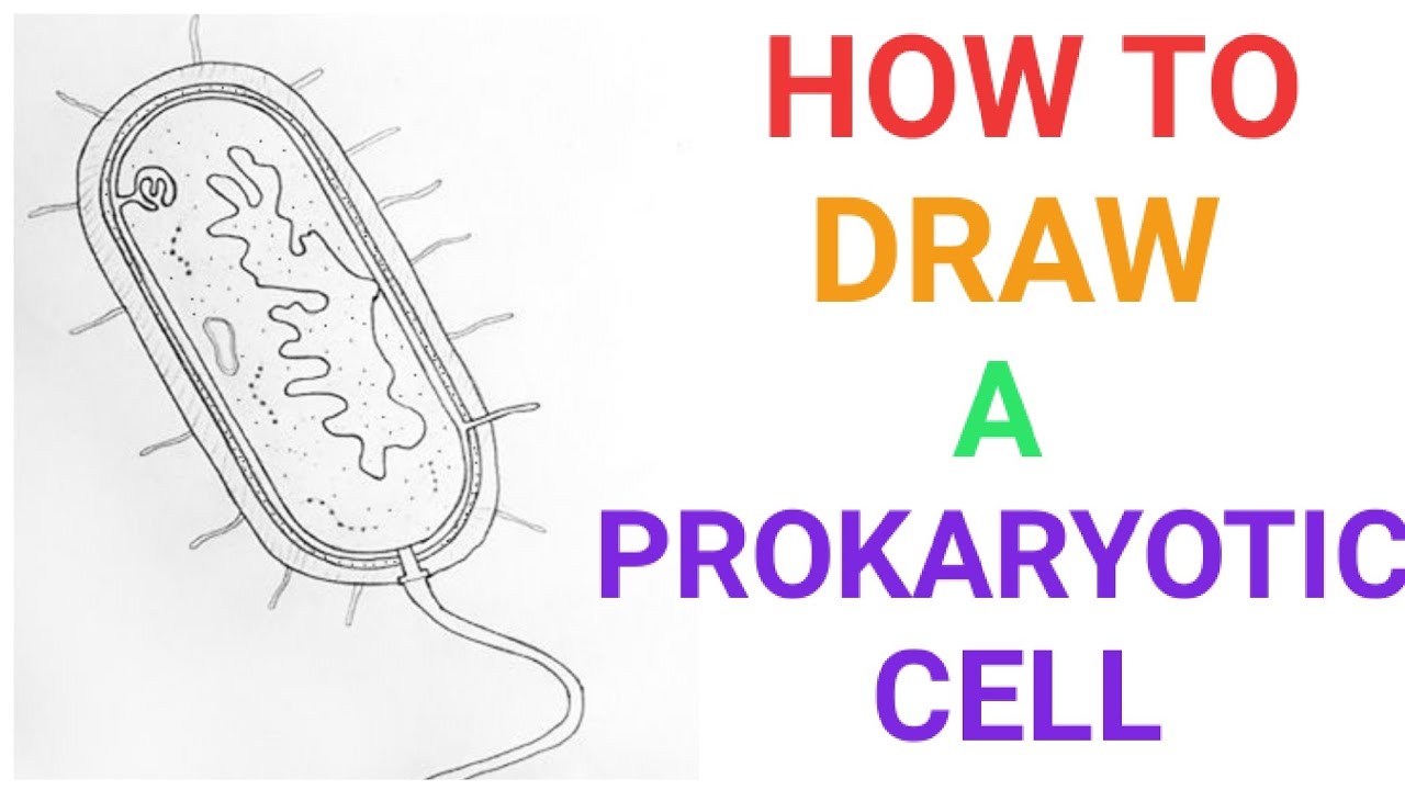
HOW TO DRAW A PROKARYOTIC CELL. YouTube
How to draw a prokaryotic cell prokaryotic organism Bacterial cell
How to draw easily PROKARYOTIC CELLS / STRUCTURE and FUNCTION / w

Draw a well labelled diagram of a prokaryotic cell.
Hello Friends!!!!In This Video, I Will Be Showing You That How To Draw A Prokaryotic.
Web Prokaryotic Cells 2.2.1 Draw And Label A Diagram Of The Ultrastructure Of Escherichia Coli (E.
Identify Each Of These Parts In The Diagram.
The Figure Below Shows The Sizes Of Prokaryotic, Bacterial, And Eukaryotic, Plant And Animal, Cells As Well As Other Molecules And Organisms On A Logarithmic.
Related Post: