How To Draw A Vbg
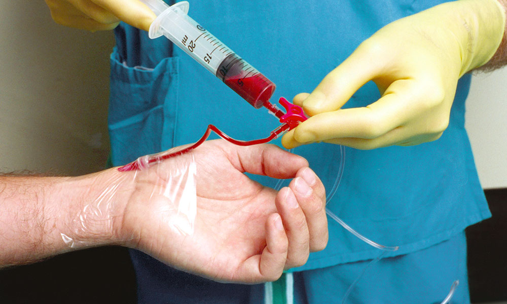
How To Draw A Vbg - The radial artery on the wrist is most commonly used to obtain the sample. Performing a vbg rather than an abg is particularly convenient in the intensive care unit and in the emergency department, either peripherally or from a central venous catheter from which venous. Pco2 is also used to track hypercarbia, as in. Web you can either use winter’s formula (expected paco2 = (1.5 x serum hco3)+ (8±2)) or expected paco2 = last two digits of the ph ± 2. Web peripheral vbg is a more humane and convenient test: Immediately place syringe on wet ice. Remove the needle, cap tightly and. Web as such, a venous blood gas (vbg) is an alternative method of estimating ph and other variables. Peripheral veins, typically the antecubital veins, are the usual sites for venous blood sampling. Once the puncture has been performed or the line specimen drawn, immediately remove all air from the syringe. Follow these procedures for blood gas sample collection and handling, sample mixing and transport to help optimize your clinical operations. Vbg may be measured along with routine venous phlebotomy for other labs. Web a venous blood gas (vbg) is an alternative method of estimating systemic carbon dioxide and ph that does not require arterial blood sampling. Web you can either. Vbg may be measured along with routine venous phlebotomy for other labs. Immediately place syringe on wet ice. Web by far the most common venous blood sample is that obtained by needle puncture of a peripheral (superficial) vein in the forearm (at the antecubital fossa) or back of the hand; In venous blood sampling, a needle is inserted into a. Web instruct patients to look away from the equipment and the procedure to help prevent a vasovagal episode. It's hard to draw any conclusions from this fact alone, so its worth looking into who owns those private companies. Captures blood from the superior and inferior vena cavae and the coronary sinus to reflect a true mixture of all of the. Web venous blood gas (vbg) analysis is a safer procedure and may be an alternative for abg. Web a venous blood gas (vbg) is an alternative method of estimating systemic carbon dioxide and ph that does not require arterial blood sampling. Make sure your creative phlebotomists don't make this mistake, too. It's hard to draw any conclusions from this fact. Peripheral veins, typically the antecubital veins, are the usual sites for venous blood sampling. Web introduction and background. Once the puncture has been performed or the line specimen drawn, immediately remove all air from the syringe. Web by far the most common venous blood sample is that obtained by needle puncture of a peripheral (superficial) vein in the forearm (at. In venous blood sampling, a needle is inserted into a vein to collect a sample of blood for testing. Make sure your creative phlebotomists don't make this mistake, too. This study was aimed to investigate the correlation of ph, pco 2, bicarbonate, sodium, potassium, and chloride (electrolytes) between abg and central vbg in icu patients. Peripheral veins, typically the antecubital. Web a vbg is a venous blood sample drawn into an abg ( heparinised) syringe and then run through a blood gas analyser. Vbg may be measured along with routine venous phlebotomy for other labs. Pco2 is also used to track hypercarbia, as in. Make sure your creative phlebotomists don't make this mistake, too. Web this video demonstrates one way. Web a venous blood gas (vbg) is an alternative method of estimating systemic carbon dioxide and ph that does not require arterial blood sampling. Web by far the most common venous blood sample is that obtained by needle puncture of a peripheral (superficial) vein in the forearm (at the antecubital fossa) or back of the hand; Web how to draw. Web how to draw an abg. Blood can be drawn via an arterial stick from the wrist, groin, or above the elbow. Web a true mixed venous sample (called svo2) is drawn from the tip of the pulmonary artery catheter, and includes all of the venous blood returning from the head and arms (via superior vena cava), the gut and. Peripheral veins, typically the antecubital veins, are the usual sites for venous blood sampling. Web peripheral vbg is a more humane and convenient test: It's hard to draw any conclusions from this fact alone, so its worth looking into who owns those private companies. Web this order is for venous blood gas for a specimen drawn from a central catheter. Peripheral iv lines can often be used to draw back a small sample of venous blood for analysis (again, without requiring a separate puncture). Remove the needle, cap tightly and. Make sure your creative phlebotomists don't make this mistake, too. Web how to draw an abg. Captures blood from the superior and inferior vena cavae and the coronary sinus to reflect a true mixture of all of the venous blood coming back to the right side of the heart. This study was aimed to investigate the correlation of ph, pco 2, bicarbonate, sodium, potassium, and chloride (electrolytes) between abg and central vbg in icu patients. Web by far the most common venous blood sample is that obtained by needle puncture of a peripheral (superficial) vein in the forearm (at the antecubital fossa) or back of the hand; Web you can either use winter’s formula (expected paco2 = (1.5 x serum hco3)+ (8±2)) or expected paco2 = last two digits of the ph ± 2. Web a true mixed venous sample (called svo2) is drawn from the tip of the pulmonary artery catheter, and includes all of the venous blood returning from the head and arms (via superior vena cava), the gut and lower extremities (via the inferior vena cava) and the coronary veins (via the coronary sinus). Web peripheral vbg is a more humane and convenient test: Web a vbg is a venous blood sample drawn into an abg ( heparinised) syringe and then run through a blood gas analyser. Follow these procedures for blood gas sample collection and handling, sample mixing and transport to help optimize your clinical operations. Vbg may be measured along with routine venous phlebotomy for other labs. Once the puncture has been performed or the line specimen drawn, immediately remove all air from the syringe. It's hard to draw any conclusions from this fact alone, so its worth looking into who owns those private companies. This is peripheral venous blood and must be distinguished.
How to Draw an Arterial Blood Gas 9 Steps (with Pictures)

Venous Blood Gas Chart

Arterial Blood Gas Interpretation Chart

How to Draw an Arterial Blood Gas 9 Steps (with Pictures)

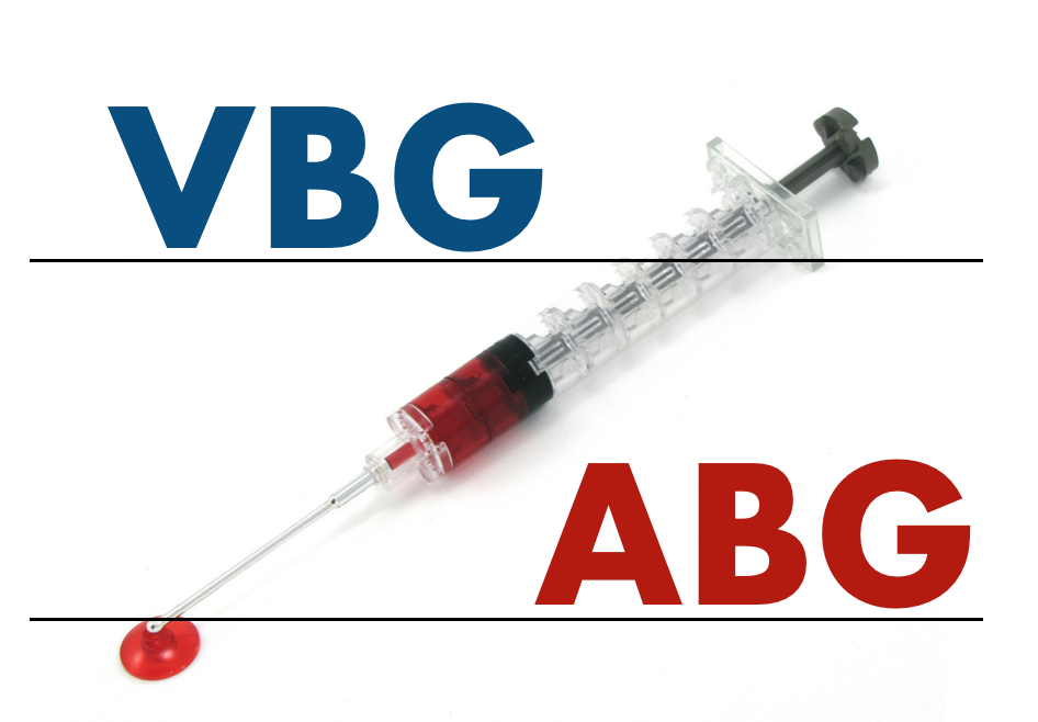
What's in a Blood Gas? VBG vs ABG — Taming the SRU

Simplified Arterial Blood Gases Simplified Nursing

How to draw and arterial blood gas YouTube
Blood Gas, Venous (VBG), Blood* Rutland Regional Medical Center

How to Draw an Arterial Blood Gas 9 Steps (with Pictures)

Basic Arterial Blood Gas Interpretation
Web A Venous Blood Gas (Vbg) Is An Alternative Method Of Estimating Systemic Carbon Dioxide And Ph That Does Not Require Arterial Blood Sampling.
Performing A Vbg Rather Than An Abg Is Particularly Convenient In The Intensive Care Unit And In The Emergency Department, Either Peripherally Or From A Central Venous Catheter From Which Venous.
Immediately Place Syringe On Wet Ice.
Web We've Heard Of Phlebotomists Drawing Blood Gases (Venous And Arterial) Into Heparinized Tubes Instead Of Blood Gas Syringes.
Related Post: