Plasma Membrane Drawing Labeled
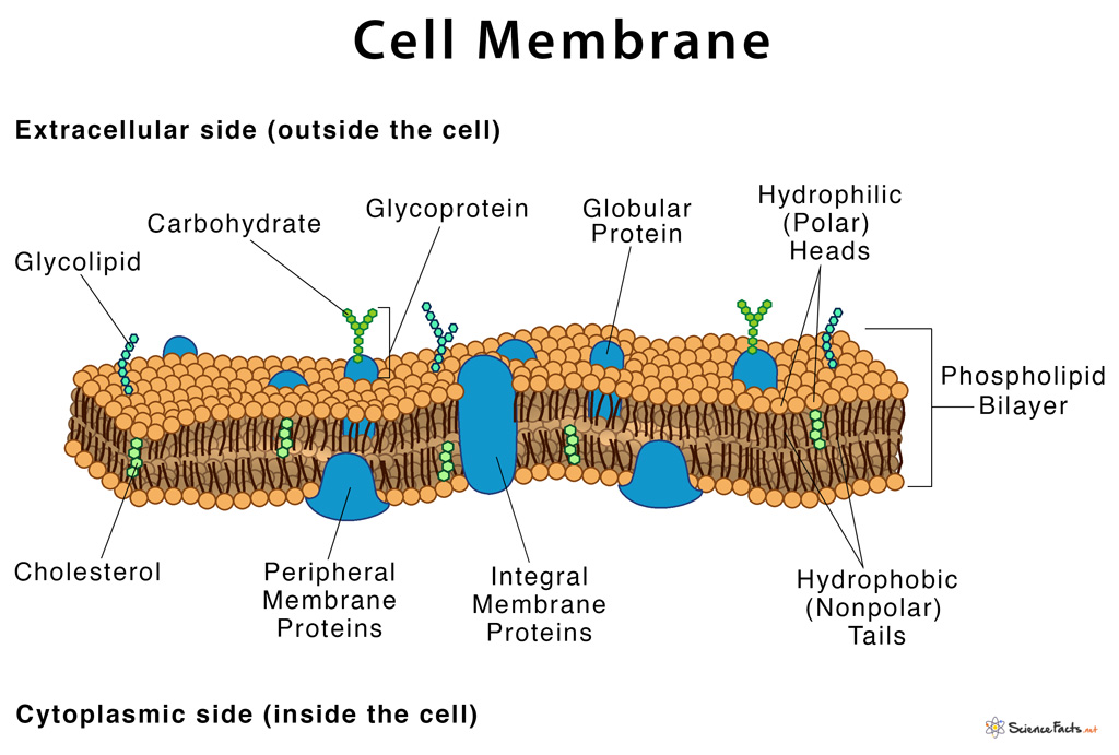
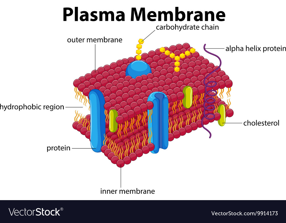
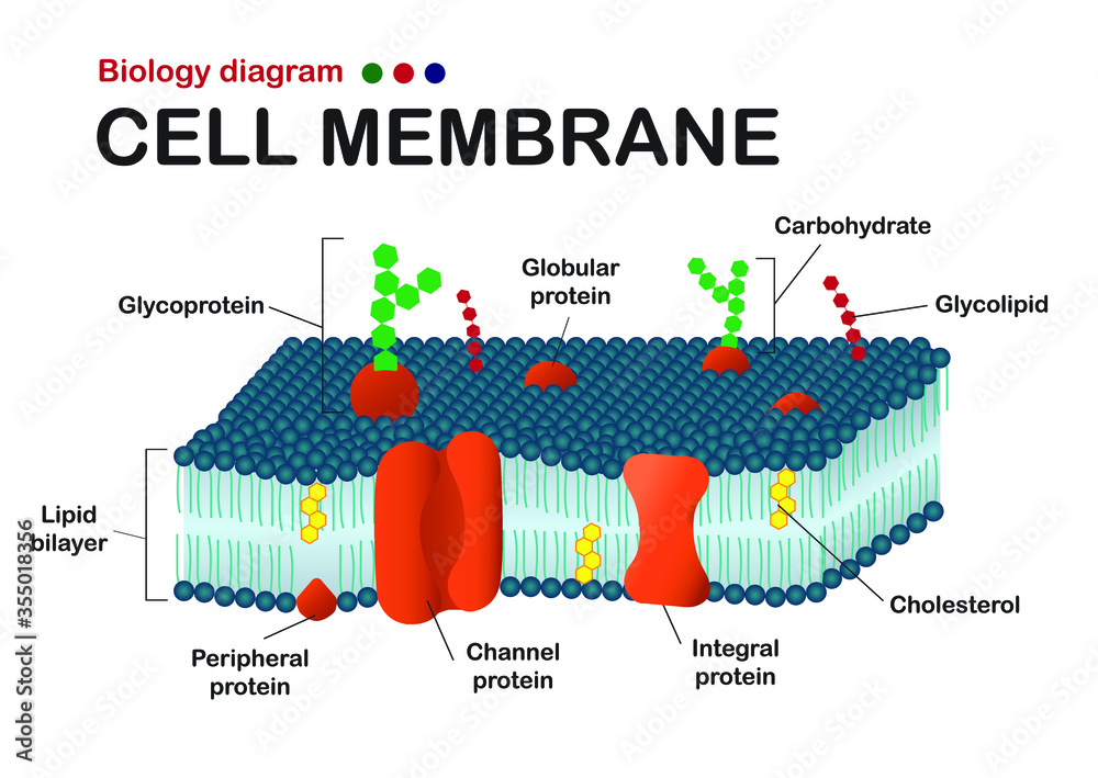
Plasma Membrane Drawing Labeled - As a comparison, human red blood cells, visible via light microscopy, are approximately 8 μm thick, or approximately 1,000 times thicker than a plasma membrane. The fluid mosaic model of the plasma membrane structure describes the plasma membrane as a fluid combination of. Web the plasma membrane—the outer boundary of the cell—is the bag, and the cytoplasm is the goo. The 3 proteins have lines with the label integral membrane proteins. Web there are two important parts of a phospholipid: In eukaryotic cells, the plasma membrane surrounds a cytoplasm filled with ribosomes and organelles. Specialized structure that surrounds the cell and its internal environment; Web peripheral protein, or peripheral membrane proteins, are a group of biologically active molecules formed from amino acids which interact with the surface of the lipid bilayer of cell membranes. Through these processes, the cell membrane is constantly renewing and changing as needed by the cell. In the case of the plasma membrane, these compartments are the inside and the outside of the cell. Web the cell membrane, also known as the plasma membrane, is a double layer of lipids and proteins that surrounds a cell. It is a sterol, a type of lipid. Both prokaryotic and eukaryotic cells have. The main function of the plasma membrane is to protect the cell from its surrounding environment. The head and the two tails. Web there are two important parts of a phospholipid: On the inner side of the phospholipid bilayer is another protein that is positioned up against the inner portion of the bilayer. Exocytosis is much like endocytosis in reverse. Web the model describes plasma membrane structure as a mosaic of components which includes proteins, cholesterol, phospholipids, and carbohydrates; Organelles are structures. Some organelles (nuclei, mitochondria, chloroplasts) are even surrounded by double membranes. It is also simply called the cell membrane. In the case of the plasma membrane, these compartments are the inside and the outside of the cell. The illustration of the plasma membrane is from your lesson. It is a feature of all cells, both prokaryotic and eukaryotic. A 3d diagram of the cell membrane. Function of the cell membrane. Exocytosis is much like endocytosis in reverse. It is also simply called the cell membrane. Web the cell membrane, also known as the plasma membrane, is a double layer of lipids and proteins that surrounds a cell. It is a selectively permeable cell organelle,allowing certain substances inside the cell while preventing others to pass through and thus is analogous to a barrier or gatekeeper. A 3d diagram of the cell membrane. The enclosure provided by the plasma membrane protects cells from their environment both mechanically and chemically. The head and the two tails. In the case of. Through these processes, the cell membrane is constantly renewing and changing as needed by the cell. Cell chemistry & cell components biological membranes. In the case of the plasma membrane, these compartments are the inside and the outside of the cell. Protein type that spans the plasma membrane activity 2: It imparts a fluid character on the membrane. Web a cell’s plasma membrane defines the boundary of the cell and determines the nature of its contact with the environment. Web the addition of new membrane to the plasma membrane is usually coupled with endocytosis so that the cell is not constantly enlarging. Web membranes and their components have the following functions: The illustration of the plasma membrane is. One of the proteins is shown with a channel in it. The head is a phosphate molecule that is attracted to water (hydrophilic).the two tails are made up of fatty acids (chains of carbon atoms) that aren’t compatible with, or repel, water (hydrophobic).the cell membrane is exposed to water mixed with electrolytes and other. Cell chemistry & cell components biological. Web the cell membrane, also called the plasma membrane, is a thin layer that surrounds the cytoplasm of all prokaryotic and eukaryotic cells, including plant and animal cells. The enclosure provided by the plasma membrane protects cells from their environment both mechanically and chemically. The fundamental structure of the membrane is the phospholipid bilayer, which forms a stable barrier between. The illustration of the plasma membrane is from your lesson. Both prokaryotic and eukaryotic cells have. The enclosure provided by the plasma membrane protects cells from their environment both mechanically and chemically. Specialized structure that surrounds the cell and its internal environment; Plasma membranes enclose the borders of cells, but rather than being a static bag, they are dynamic and. Web the fluid mosaic model describes the plasma membrane structure as a mosaic of components—including phospholipids, cholesterol, proteins, and carbohydrates—that gives the membrane a fluid character. Some organelles (nuclei, mitochondria, chloroplasts) are even surrounded by double membranes. It separates the cytoplasm (the contents of the cell) from the external environment. For comparison, human red blood cells, visible via light microscopy, are approximately 8. Controls movement of substances into/out of cell. Molecule that contains both a hydrophobic and a hydrophilic end. Web membranes and their components have the following functions: Web a diagram of a plasma membrane shows a phospholipid bilayer with 3 proteins embedded in the bilayer. The plasma membrane mediates cellular processes by regulating the materials that enter and exit. One of the proteins is shown with a channel in it. Enclosure and insulation of cells and organelles. Exocytosis is much like endocytosis in reverse. Of course, a cell is ever so much more than just a bag of goo. Protein type that spans the plasma membrane activity 2: Specialized structure that surrounds the cell and its internal environment; Learn vocabulary, terms, and more with flashcards, games, and other study tools.
Labeled Diagram Of Plasma Membrane Best Of Plasma Membrane Diagrams

Labelled Diagram Of Cell Membrane

5.4 Plasma Membrane Biology LibreTexts

Plasma Membrane Diagrams 101 Diagrams

Diagram with plasma membrane Royalty Free Vector Image

Plasma Membrane Diagrams 101 Diagrams

Plasma membrane. Molecular structure of plasma membrane, eps8 , Ad,

STRUCTURE of PLASMA MEMBRANE

Biology diagram show structure of cell membrane (or plasma membrane
:max_bytes(150000):strip_icc()/cell-membrane-373364_final-5b5f300546e0fb008271ce52.png)
Cell Membrane Function and Structure
The Proportion Of Constituency Of Plasma Membrane I.e., The Carbohydrates, Lipids And Proteins Vary.
Plasma Membranes Range From 5 To 10 Nm In Thickness.
Web The Plasma Membrane Of A Cell Is A Network Of Lipids And Proteins That Forms The Boundary Between A Cell’s Contents And The Outside Of The Cell.
Web There Are Two Important Parts Of A Phospholipid:
Related Post: