Typical Human Cell Drawing
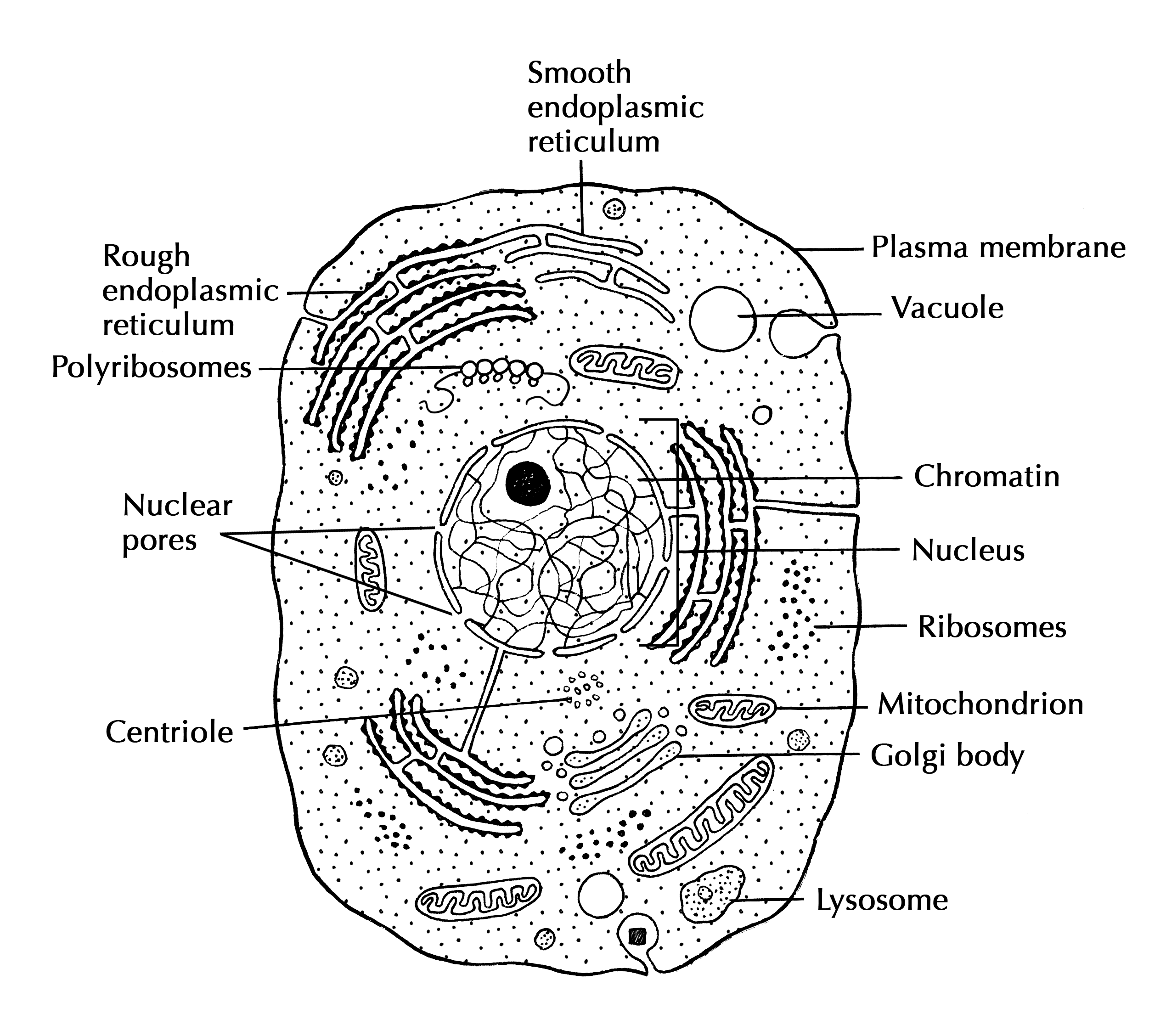
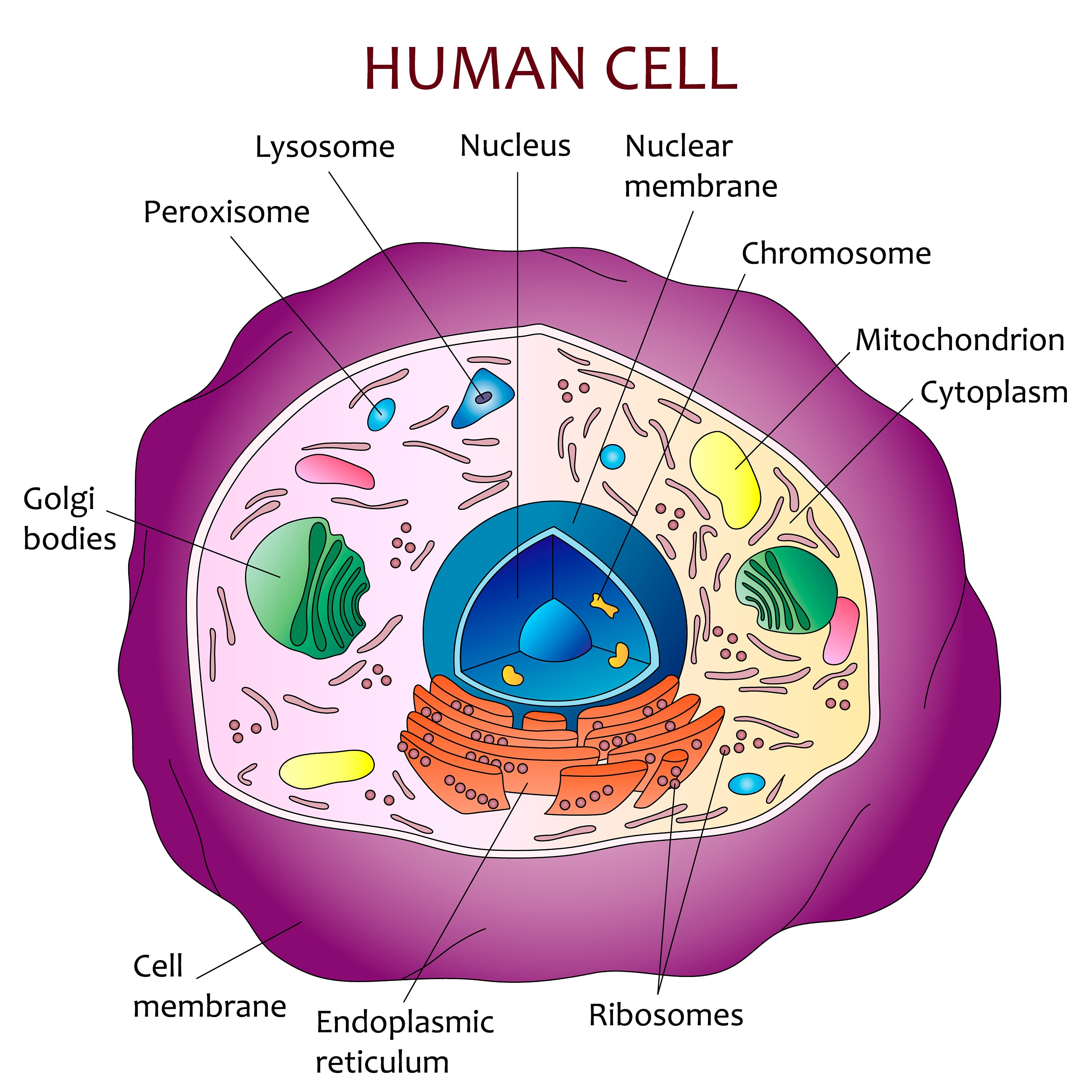
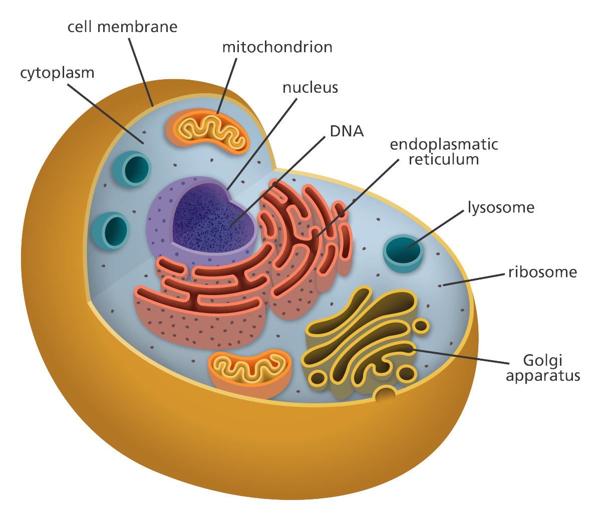
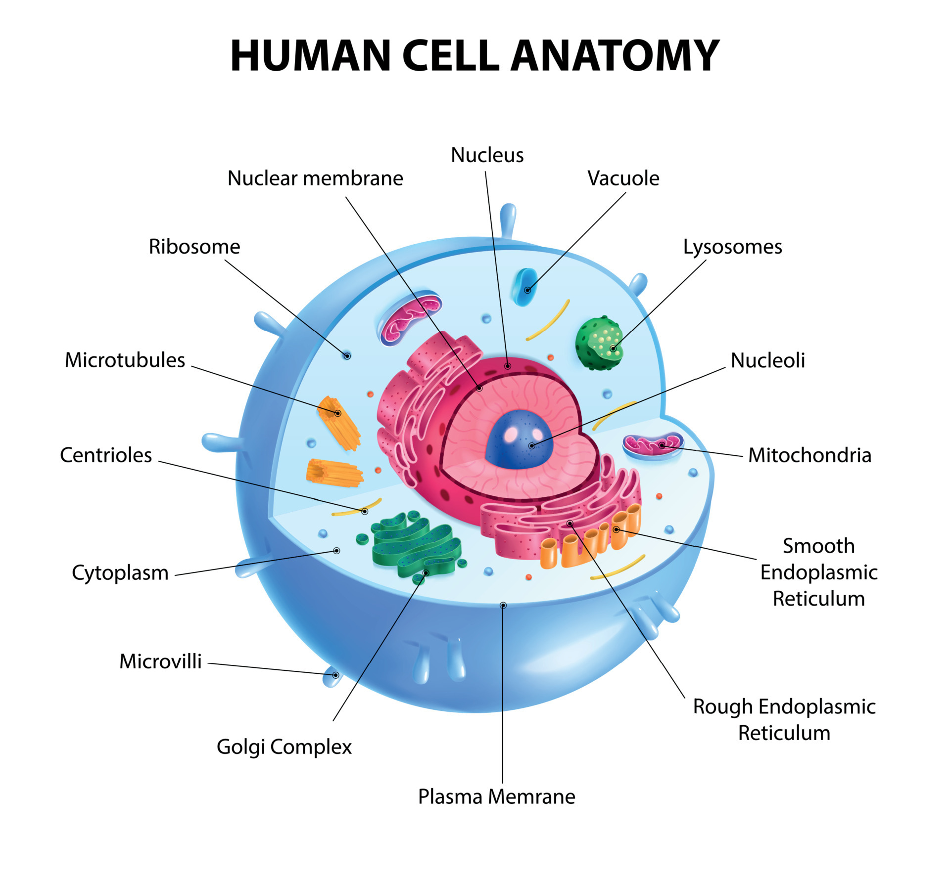
Typical Human Cell Drawing - Table \(\pageindex{1}\) below describes the functions of mitochondrion, rough and smooth endoplasmic reticulum, golgi apparatus, secretory vesicles, peroxisomes, lysosomes, microtubules and microfilaments (fibers of the. Together, trillions of cells make up the human body. A single cell may be a complete organism in itself, such as a bacterium, or it may acquire a specialized function, becoming a building block of a multicellular organism. The cell membrane is the outer coating of the cell and contains the cytoplasm, substances within it and the organelle. Why don’t we take a look at the inside of a typical cell? The cell membrane is the outer coating of the cell and contains the cytoplasm, substances within it and the organelle. Web interactive guide to stem cells and cell biology with 3d models and real microscopy data of gfp labeled hipscs. Also know that the membrane is not a rigid cell wall like in plant cells. Web diagram of the human cell illustrating the different parts of the cell. Web we will cover a large number of subcellular structures that are unique to eukaryotes, and you will certainly be expected to know the names of these structures or organelles, to associate them with one or more functions, and to identify them on a canonical cartoon representation of a eukaryotic cell. Eukaryotic cell diagram, vector illustration, text on own layer. The important part is that it does not have any sharp edges. Web get animal cell facts, including a labeled cell diagram, a list of organelles and their functions, and a summary of animal cell types. Web what is your request drawing?please comment below. An animal cell is a eukaryotic cell. Eukaryotic cell diagram, vector illustration, text on own layer. Unlike the animal cell lacking the cell wall. The plasma membrane is exactly what it sounds like: The cell membrane is the outer coating of the cell and contains the cytoplasm, substances within it and the organelle. The cytoplasm is the region outside the nucleus that contains cell organelles and cytosol,. Eukaryotic cell diagram, vector illustration, text on own layer. Web diagram of the human cell illustrating the different parts of the cell. The interior of human cells is divided into the nucleus and the cytoplasm. You can make the circle misshapen or oblong. Diagram of the human cell illustrating the different parts of the cell. Web diagram of the human cell illustrating the different parts of the cell. There exist two general classes of cells: Web cell diagrams showing a typical animal cell and plant cell. Unlike the animal cell lacking the cell wall. Cross section animal cell structure detailed colorful anatomy. Learn vocabulary, terms, and more with flashcards, games, and other study tools. Eukaryotic cell diagram, vector illustration, text on own layer. Together, trillions of cells make up the human body. Web get animal cell facts, including a labeled cell diagram, a list of organelles and their functions, and a summary of animal cell types. The interior of human cells is. Web figure \(\pageindex{5}\) typical example of a cell containing the primary organelles and internal structures. You can make the circle misshapen or oblong. The cell membrane is the outer coating of the cell and contains the cytoplasm, substances within it and the organelle. The cell membrane is the outer coating of the cell and contains the cytoplasm, substances within it. Cross section animal cell structure detailed colorful anatomy. The cell organelles are enclosed by the plasma membrane including the cell nucleus. Web the genetic information within each cell acts as a sort of instruction manual, telling a cell how to function and replicate. The cytoplasm is the region outside the nucleus that contains cell organelles and cytosol, or cytoplasmic solution.. :)thanks for watching our channel. Web a cell is the smallest living organism and the basic unit of life on earth. Cross section animal cell structure detailed colorful anatomy. There exist two general classes of cells: 577 views 2 years ago #drawing #pencil #easy. The cell membrane is the outer coating of the cell and contains the cytoplasm, substances within it and the organelle. Web get animal cell facts, including a labeled cell diagram, a list of organelles and their functions, and a summary of animal cell types. The difference is simple and readily recognizable under light microscopy. Web what is your request drawing?please. Web start studying typical human cell. It provides a clear view of the structure and organization of the cell, allowing scientists and students to study and identify the different components. 577 views 2 years ago #drawing #pencil #easy. Web figure \(\pageindex{5}\) typical example of a cell containing the primary organelles and internal structures. Draw a simple circle or oval for. Cross section animal cell structure detailed colorful anatomy. Web figure \(\pageindex{5}\) typical example of a cell containing the primary organelles and internal structures. 577 views 2 years ago #drawing #pencil #easy. Also know that the membrane is not a rigid cell wall like in plant cells. Web there are two basic types of cells: Web diagram of the human cell illustrating the different parts of the cell. The difference is simple and readily recognizable under light microscopy. The plasma membrane is exactly what it sounds like: Web get animal cell facts, including a labeled cell diagram, a list of organelles and their functions, and a summary of animal cell types. In humans, as in all organisms, cells perform all functions of life. 4.9k views 11 months ago india. The interior of human cells is divided into the nucleus and the cytoplasm. A membrane made of plasma. Unlike the animal cell lacking the cell wall. Web the human cell diagram unlabeled is a visual representation of the different parts of a typical human cell without any labels or descriptions. Table \(\pageindex{1}\) below describes the functions of mitochondrion, rough and smooth endoplasmic reticulum, golgi apparatus, secretory vesicles, peroxisomes, lysosomes, microtubules and microfilaments (fibers of the.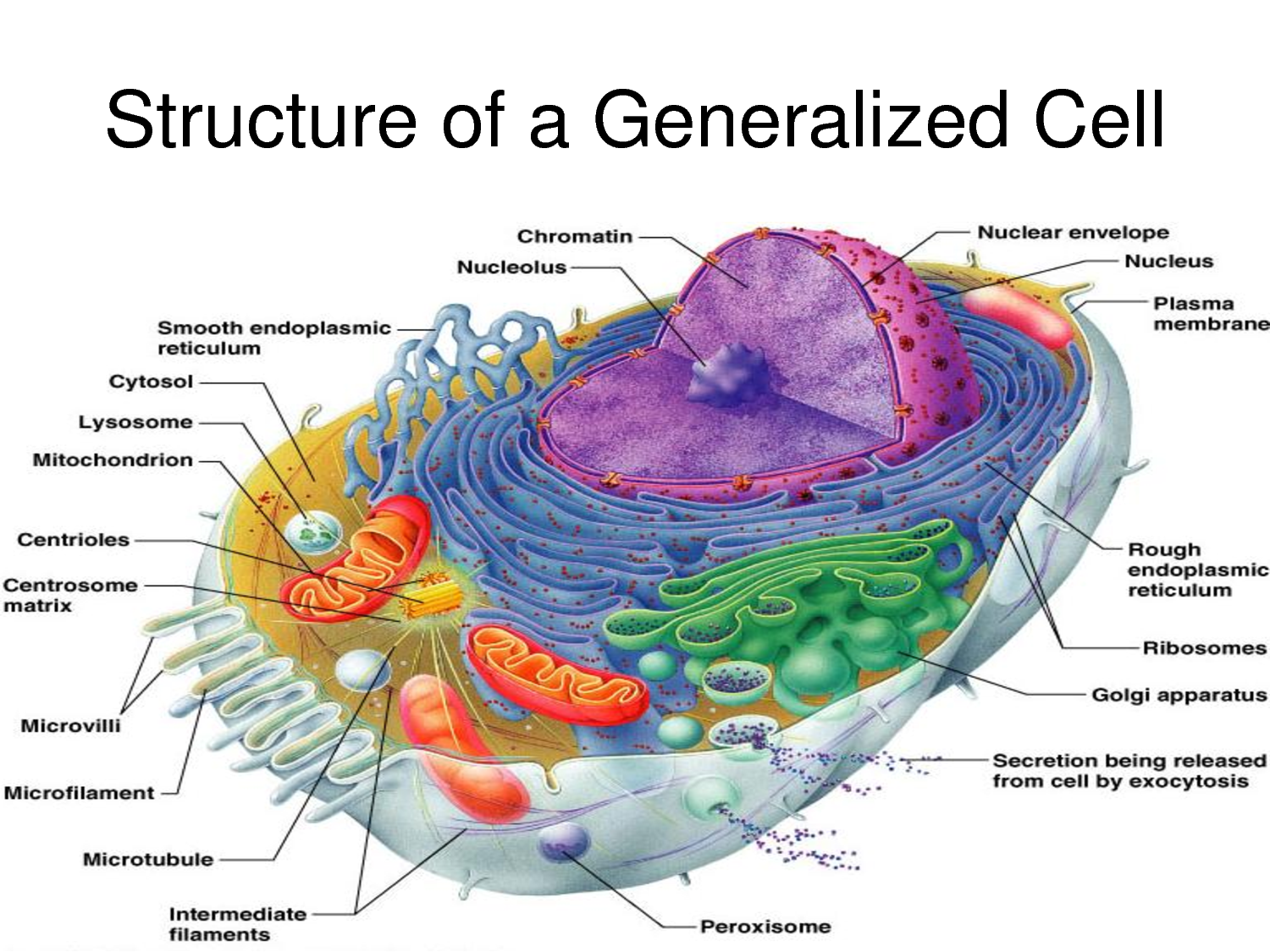
Human Cell Diagrams Images & Pictures Becuo

Blood Cell Organelles Cell Organelles

Biochemistry Lecture

Human Cell Diagram, Parts, Pictures, Structure and Functions
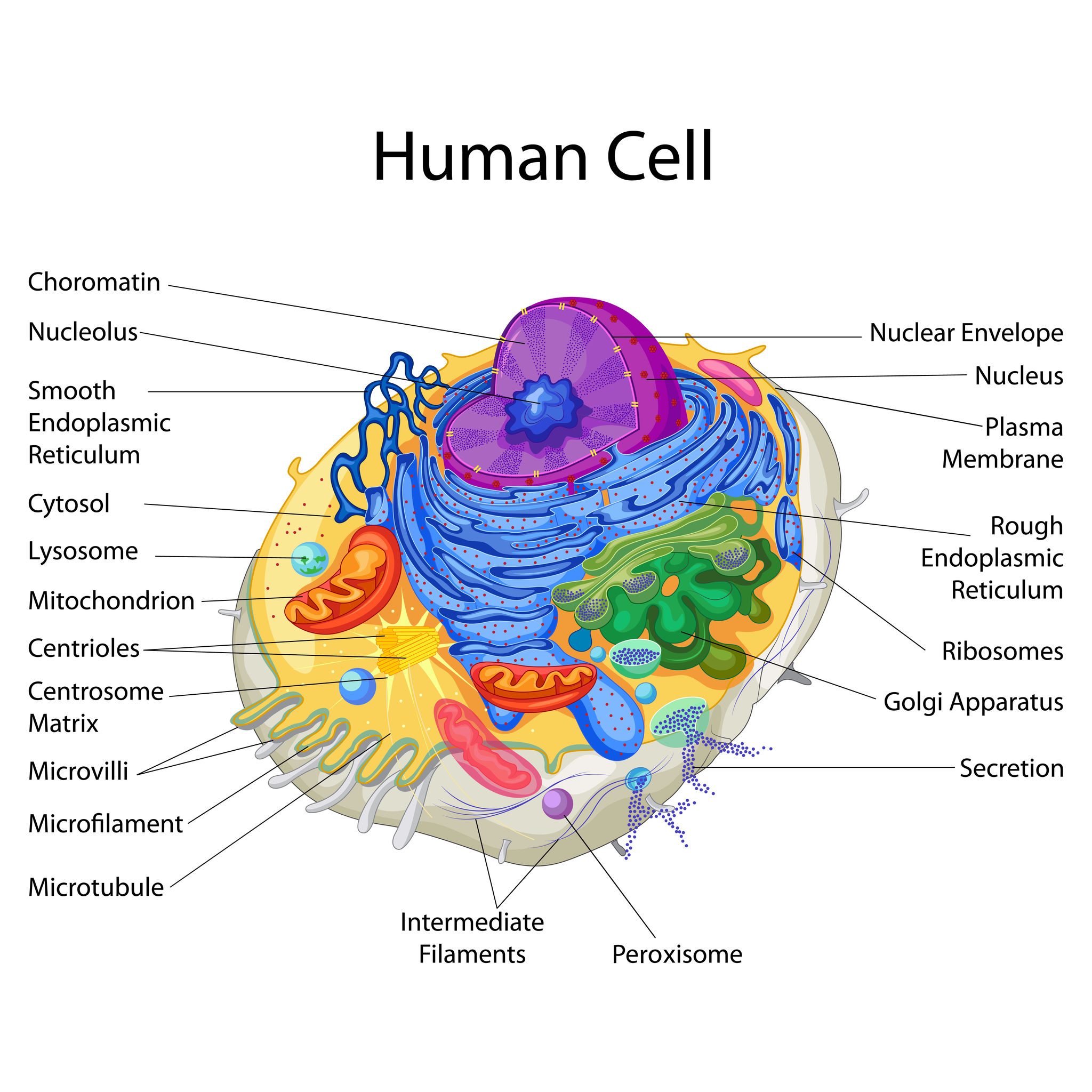
diagram of a human cell
Human Cell Sketch at Explore collection of Human
Human cell diagram Etsy
Human Cell Anatomy Diagram

How to Draw Human Cell Step by Step YouTube
Human Cell Diagram 6406474 Vector Art at Vecteezy
The Cell Organelles Are Enclosed By The Plasma Membrane Including The Cell Nucleus.
Web Interactive Guide To Stem Cells And Cell Biology With 3D Models And Real Microscopy Data Of Gfp Labeled Hipscs.
The Membrane, The Nucleus, And The Cytoplasm.
Structure, Parts, Functions, Labeled Diagram.
Related Post: